Farm & Ranch
Avian Influenza Update
Barry Whitworth, DVM
Area Food/Animal Quality and Health
Specialist for Eastern Oklahoma
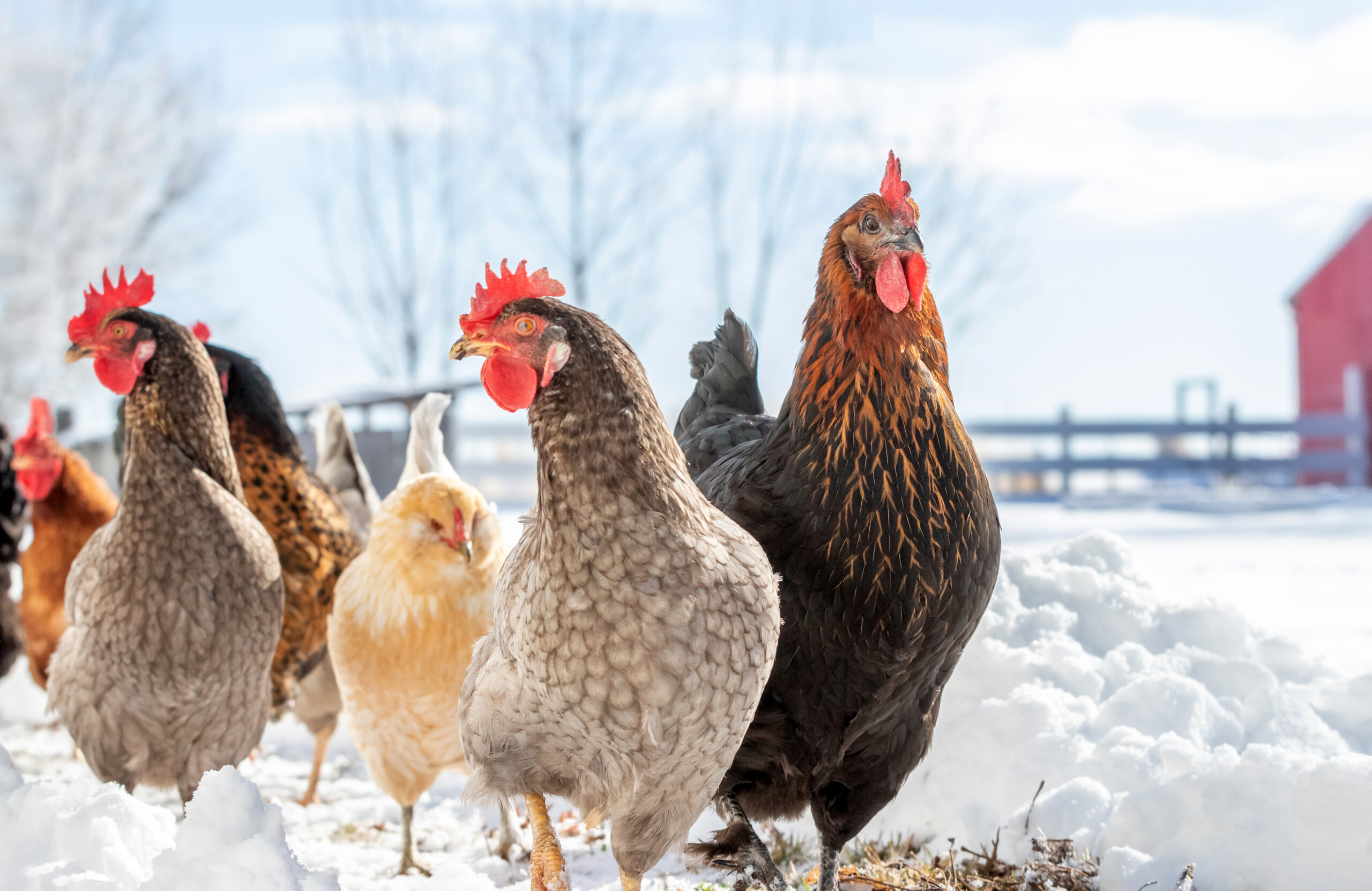
High Path Avian Influenza (HPAI) continues to be a problem in commercial and backyard poultry in the Unites States (US) with over 60 million birds affected. Since the start of the outbreak in 2022, 879 flocks (347 commercial and 532 backyard flocks) have been confirmed with HPAI in the US. Many wild birds and mammals have been affected as well. Five backyard flocks and one commercial flock have been confirmed with HPAI during this outbreak in Oklahoma. The latest was detected in a backyard flock in Carter County on October 16, 2023. For a complete listing of domestic birds, wild birds, and mammals affected by HPAI visit 2022-2023 Detection of High Path Avian Influenza website at https://www.aphis.usda.gov/aphis/ourfocus/animalhealth/animal-disease-information/avian/avian-influenza/2022-hpai.
Avian influenza (AI) is a highly contagious viral disease. The virus is classified as either Low Path Avian Influenza (LPAI) or HPAI depending on the virulence. This virus infects many food producing birds such as chickens and turkeys while it commonly resides in migratory waterfowl and many other wild birds. Most often ducks, geese, and wild birds harbor the virus in the intestinal tract without having any clinical signs of the disease. The virus is shed in the feces and respiratory secretions from infected birds. Poultry can be infected with the virus when they come in direct contact with infected birds or consume feed that is contaminated with the virus. The virus can be spread indirectly through objects like shoes, clothes, or equipment contaminated with the virus.
Clinical signs of the disease vary depending on the severity of the virus and the organ system affected. LPAI usually results in no clinical signs or only mild problems. However, HPAI has many different clinical signs. Death with no symptoms is a common finding. Respiratory problems such as coughing, sneezing, watery eyes, and nasal discharges may be seen. Depression resulting in loss of appetite and decrease consumption of water may occur. Egg production may be impacted with a decrease in production and/or softshell or misshapen eggs. A bird’s comb, wattle, head, eyelids, and hocks may swell. Combs and wattles may turn purple. Nervous system disorders including tremors, incoordination, and unusual positions of the head may be seen. Diarrhea has been reported in some cases. For more information about clinical signs visit Defend the Flock-Signs of Illness at https://www.aphis.usda.gov/aphis/ourfocus/animalhealth/animal-disease-information/avian/defend-the-flock-program/outbreak-illness/outbreak-illness.
For commercial and backyard poultry flocks, the best defense against HPAI is a sound biosecurity program. Biosecurity is the development and implementation of management procedures intended to reduce or prevent unwanted threats from entering a flock. The protocol is designed to reduce or prevent the spread of unwanted threats through the flock and eliminate any unwanted pathogens that may enter the flock. Lastly, a biosecurity plan is designed to prevent threats from infecting neighboring poultry operations. Biosecurity can be broken down into four basic areas which include traffic, isolation, sanitation, and husbandry.
The first line of defense should be limiting the traffic that enters the area. Poultry operations should have a perimeter buffer area (PBA). For backyard poultry operations, this could be a fence. In commercial operations this may be a fence or road that surrounds the facility. All entry points need to be clearly marked with “Do Not Enter” signs. In a study by United States Department of Agriculture (USDA) evaluating factors associated with introduction of HPAI in layer farms in the US, the presence of a gate was found to be protective against the introduction of the virus. Gates with signage may encourage people to follow biosecurity protocols.
Inside the PBA, a line of separation (LOS) needs to be established. The LOS isolates the birds from possible sources of infections. The LOS is usually the walls of the poultry building plus the entry point. No person should cross this line without following proper biosecurity protocols. Producers should provide visitors with clean coveralls and disposable shoe covers. Visitors should wash their hands before and after visiting the facility. All visitors should dip their shoes in a disinfectant solution when entering and exiting the facility. Also, no other animals, wild or domestic should cross the LOS.
Sanitation is one of the most important parts of a biosecurity plan. All equipment, feeders, waterers, and buildings need to be cleaned and disinfected regularly. First, all fecal material and dirt should be physically removed. Next, disinfectants must be applied and allowed sufficient contact time to work properly. Foot baths need to be properly maintained. The property outside the poultry house should be kept mowed and cleaned. Failure to keep the grass cut and/or to promptly clean up feed spills is associated with HPAI.
Poultry producers must also practice good animal husbandry. Flocks need to be observed several times per day. Producers need to collect and dispose of dead birds frequently. Producer should know the clinical signs of a sick bird. Any unusual increases in sick or dead birds should be reported to proper authorities. Backyard producers have several options. They can contact their veterinarian or Oklahoma State University County Extension office. They can also contact the Oklahoma State Veterinarian at 405-522-6141.
The National Poultry Improvement Plan (NPIP) has guidelines for a biosecurity protocol. Commercial and backyard poultry producers should examine the NPIP 14 standards of the biosecurity protocol. Any areas that do not meet the standards need to be addressed. The NPIP biosecurity audit form can be found at http://www.poultryimprovement.org/documents/AuditForm-2018BiosecurityPrinciples.pdf. Additional sources for backyard poultry producers can be found at the USDA Defend the Flock website at healthybirds.aphis.usda.gov, Protect Your Poultry From Avian Influenza at https://www.aphis.usda.gov/publications/animal_health/bro-protect-poultry-from-ai.pdf or Oklahoma State University fact sheet Small Flock Biosecurity for Prevention of Avian Influenza ANSI-8301.
Avian Influenza is a major threat to the US and Oklahoma poultry industry. It is the responsibility of all commercial and backyard poultry producers to do everything in their power to protect this industry.
Reference
Swayne, D.E. and Halvorson, D.A. 2003 Influenza. In Y. M. Saif (ed.). Diseases of Poultry, 11th ed. Iowa State Press: Ames, Iowa, 135-160.
Green, A. L., Branan, M., Fields, V. L., Patyk, K., Kolar, S. K., Beam, A., Marshall, K., McGuigan, R., Vuolo, M., Freifeld, A., Torchetti, M. K., Lantz, K., & Delgado, A. H. (2023). Investigation of risk factors for introduction of highly pathogenic avian influenza H5N1 virus onto table egg farms in the United States,
2022: a case-control study. Frontiers in veterinary science, 10, 1229008.
Barry Whitworth, DVM | Senior Extension Specialist | Department of Animal & Food Sciences
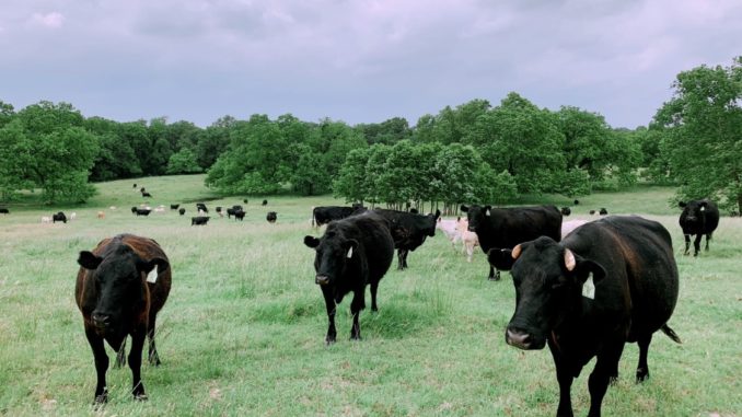
According to the Mesonet, Oklahoma received some much-needed rain in late April (2023). With the moderate temperatures and high humidity, the environment is perfect for the proliferation of gastrointestinal nematodes (GIN) which are commonly called “worms.” Cattle can be infected with a variety of GIN. Most do not cause issues unless husbandry practices are poor. However certain GIN have been associated with disease. The most pathological GIN in cattle is Ostertagia ostertagi. Cooperia species and Haemonchus species are two that have been implicated with production issues. Control of these parasites is constantly changing due to environment, anthelmintic (dewormer) resistance, and consumer preference. Cattle producers should develop a plan to manage these parasites.
In order for GIN to complete their life cycle, certain environmental conditions must exist. The development stage begins with passing of the egg in the feces of the animal. If the egg is to hatch, the temperature must be warm and the humidity needs to be close to 100%. Ideal temperature ranges from 70⁰ to 80⁰ Fahrenheit (F), but any temperature above 45⁰ F will allow for development. Temperatures above 85⁰ F or below 45⁰ F will begin to hamper development. Humidity needs to be 80% or higher.
Once the egg hatches, the larva goes through a couple of molts to reach the infective stage which is the third stage larva (L3). L3 must have moisture to free itself from the fecal pat. Once free, it rides a wave of water on to a blade of forage. Once ingested, this begins the prepatent or pre-adult stage. Two molts take place during this stage (L3 to L4 and L4 to L5). If conditions are not favorable for survivability of offspring, L4 will go into an arrested development stage (hypobiosis) for a period of time. The patent or adult stage is the mature breeding adult.
Once inside the body, the parasite will migrate to certain locations in the digestive tract. For example, O. ostertagi develop in the gastric gland in the abomasum. H. placei and H. contortus will migrate to the abomasum. Cooperia species will live in the small intestine. A few like Trichuris (whipworms) are found in the large intestine.
Clinical signs of parasitism vary according to the species of parasite, burden, and site of attachment. Severe disease, which is referred to as parasitic gastroenteritis (PGE), with internal parasites is unusual with today’s control methods. Clinical signs of PGE are lack of appetite, weight loss, weakness, diarrhea, submandibular edema (bottle jaw), and death. However, most parasite infection are subclinical which means producers do not see clinical signs of disease. In subclinical infections, the parasite causes production issues such as poor weight gain in young cattle, reduced milk production, and lower pregnancy rates.
Producers should be monitoring their herds for parasites throughout the year but especially in the spring when conditions are ideal for infection. A fecal egg count (FEC) is a good way of accessing parasite burdens. Livestock producers need to gather fecal samples from their herd periodically. The samples should be sent to their veterinarian or a veterinary diagnostic lab. Different techniques are used to access the number of eggs per gram of feces. Based on the counts, the producer will learn the parasite burden of the herd. Producers can use this information to develop a treatment plan.
In the past, GIN control was simple. Cattle were routinely dewormed. Unfortunately, anthelmintic resistance has complicated parasite control. Now proper nutrition, grazing management, a general understanding of how weather influences parasites, biosecurity, refugia, anthelmintic efficiency, and the judicious use of anthelmintics are important in designing an effective parasite management program. All of these considerations need to be discussed in detail with a producer’s veterinarian when developing a plan for their operation.
Cattle producers need to understand that parasites cannot be eliminated. They must be managed with a variety of control methods. Designing a parasite management plan requires producers to gain a general understanding of life cycle of the parasite as well as the environmental needs of the parasite. Producers should use this information as well as consult with their veterinarian for a plan to manage GIN. For more information about GIN, producers should talk with their veterinarian and/or with their local Oklahoma State University Cooperative Extension Agriculture Educator.
References
Charlier, J., Höglund, J., Morgan, E. R., Geldhof, P., Vercruysse, J., & Claerebout, E. (2020). Biology and Epidemiology of Gastrointestinal Nematodes in Cattle. The Veterinary clinics of North America. Food animal practice, 36(1), 1–15.
Navarre C. B. (2020). Epidemiology and Control of Gastrointestinal Nematodes of Cattle in Southern Climates. The Veterinary clinics of North America. Food animal practice, 36(1), 45–57.
Urquhart, G. M., Armour, J., Duncan, J. L., Dunn, A. M., & Jennings, F. W. (1987). In G. M. Urquhart (Ed). Veterinary Helminthology. Veterinary Parasitology (1st ed., pp 3-33). Longman Scientific & Technical.
Barry Whitworth, DVM – Area Food/Animal Quality and Health – Specialist for Eastern Oklahoma
A ranch in Australia experienced an abnormally high number of stillbirths and weak born calves in 2004-2005. An investigation revealed that the usual infectious causes were not the problem. After additional testing, veterinarians diagnosed low levels of vitamin A as the cause.
According to Dr. Greg Hanzlicek, with the Kansas State Veterinary Diagnostic Laboratory (KSVDL), Kansas had an unusually high number of stillbirth cases and weak born calves in the spring of 2019. After many laboratory tests, it was concluded that the problem stimmed from a lack of energy, protein, Vitamin A, or combinations of all of these.
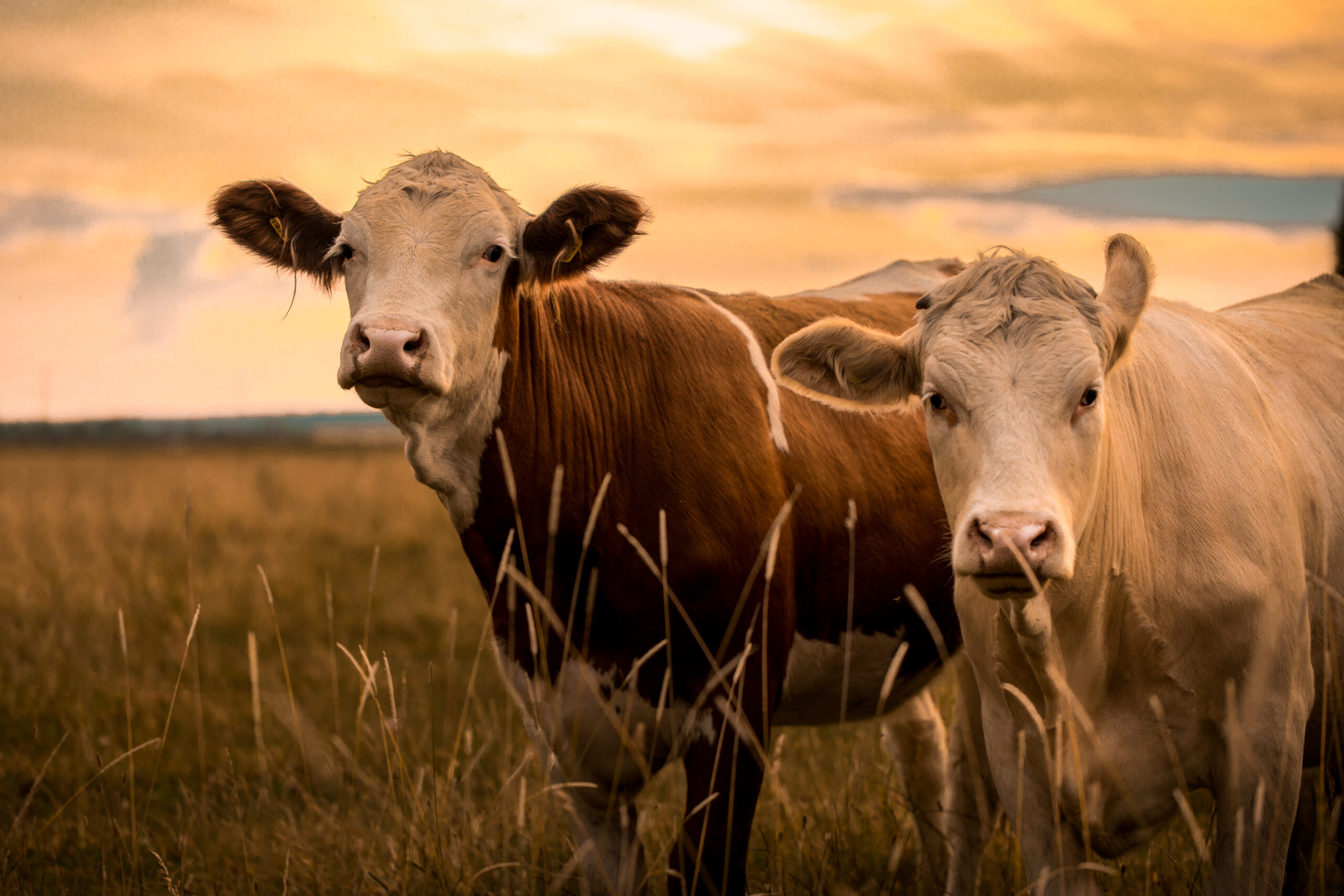
Both of the above examples demonstrate the importance of vitamin A in reproductive efficiency. Research has shown that low vitamin A levels during pregnancy are associated with abortions, stillbirths, and weak born calves. In addition to playing an important role in reproductive efficiency, vitamin A is essential for vision, bone growth, and maintaining epithelial tissue such as skin and hooves.
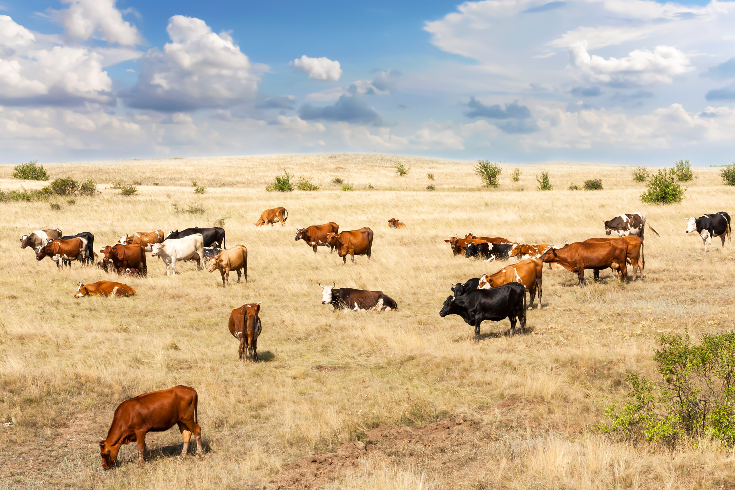
Animals obtain vitamin A from consuming green forage and/or the addition of vitamin A supplements to the diet. Lush green pastures contain high amounts of vitamin A. As plants mature and during times of drought, the amount of vitamin A decreases. The ranch in Australia experienced below average rainfall in the previous two years prior to the calving season. During the calving season, rainfall was below average with very dry conditions and little green forage was available.
In general, animals obtain adequate amounts of vitamin A by grazing green forage. Animals grazing green pastures will build a healthy store of vitamin A in the liver. When vitamin A is in short supply, the stores in the liver prevent deficiencies. According to Dr. Lalman, Extension Beef Cattle Specialist Oklahoma State University, the stores should last 2 to 4 months during times of deficiency. During times when green forage is not available, vitamin A supplements need to be added to the diet to prevent deficiencies.
When vitamin A levels are deficient, night blindness is one of the earliest clinical signs. Other eye issues include clouding of the cornea, ocular discharges, and possible ulcerations. Skin issues found when levels of vitamin A are deficient include a dry rough coat, scales on the skin, and dry cracked hooves. Other neurological signs include incoordination or gait problems. Seizures may occur due to the increase cerebrospinal fluid pressure. Birth defects have also been attributed to low vitamin A levels.
Animals displaying vitamin A deficiency should be treated immediately with vitamin A injections. If treated early, response is usually rapid and complete. However, delaying treatment may result in irreversible damage. Even with treatment, cattle with vision impairment due to vitamin A deficiency may not regain their sight.
Preventing Vitamin A deficiency depends on producers being attentive to the environmental conditions that favor low vitamin A levels in forage. During these times, producers need to supplement the diet with vitamin A. Producers need to be aware that Vitamin A supplements degrade rapidly, so vitamin A supplements should not be stored for long periods of time. In addition to vitamin A supplementation, research indicates that diets low in protein result in poor absorption of vitamin A. It is important that producers ensure that the rations have sufficient protein levels. Lastly, since colostrum contains high levels of vitamin A, producers need to ensure that newborns obtain adequate amounts of colostrum at birth.
Similar to the Australian example, most of Oklahoma had below average rainfall for the year of 2022. This resulted in pasture quality decreasing earlier than normal. Due to this year’s lack of green forage, liver stores of vitamin A may be inadequate for the animal’s needs. Producers need to ensure that the diets of their cattle have adequate amounts of vitamin A, energy, and protein. For more information about Vitamin A, producers should contact their veterinarian and/or visit with their Oklahoma State University County Ag Educator.
References
Hanzlicek, G. (2019, May). Difficult Calving Season Findings:2019. Diagnostic Insights. www.ksudl.org/resources/news/diagnostic_insights/may2019/difficult-calving-season2019.html.
Hill, B., Holroyd, R., & Sullivan, M. (2009). Clinical and pathological findings associated with congenital hypovitaminosis A in extensively grazed beef cattle. Australian Veterinary Journal, 87(3), 94–98.
Parker, E. M., Gardiner, C. P., Kessell, A. E., & Parker, A. J. (2017). Hypovitaminosis A in extensively grazed beef cattle. Australian veterinary journal, 95(3), 80–84.
Barry Whitworth, DVM, MPH | Senior Extension Specialist
Department of Animal & Food Sciences | Freguson College of Agriculture | Oklahoma State University
Cattle lice cost Oklahoma cattlemen millions of dollars each year in decreased weight gains and reduced milk production. If cattle producers have not treated their cattle for lice this fall, they need to consider what type of lice control to initiate. This is especially true for cattle producers that had problems in the previous year. Cattle producers should monitor cattle closely during the months of December, January, and February. Producers should not wait until clinical signs appear before beginning treatment.
The life cycle of the different species of cattle lice are very similar. The life cycle begins with the female louse attaching her egg to a shaft of hair. The egg will hatch as a small replica of the adult. After several molts, the adult will emerge. The cycle takes around 3 to 4 weeks to complete. These newly hatched lice will spend their entire life on the host and are host specific which means cattle cannot be infected with lice from other animals.
Small numbers of lice may be found on cattle in the summer, but high populations of lice are associated with cold weather. Since cattle tend to be in closer proximity to each other in the winter, lice can spread easily between cattle. A small percentage of cattle tend to harbor larger numbers of lice. These animals are sometimes referred to as “carrier animals”, and they may be a source for maintaining lice in the herd. As with many other diseases, stress also contributes to susceptibility and infestation.
Signs of lice infections in cattle are hair loss, unthrifty cattle, and hair on fences or other objects. If producers find these signs, they may want to check a few animals for lice. They can check for lice by parting the hair and observing the number of lice per square inch. If an animal has 1 to 5 lice per square inch, they are considered to have a low infestation. Cattle with 6 to 10 lice would be considered moderately infested. Any cattle with more than 10 lice per square inch are heavily infested.
Cattle have two types of lice. One type is the biting or chewing louse. These lice have mouth parts that are adapted to bite and chew the skin. The second type is sucking louse. These lice have mouth parts that will penetrate the skin and suck blood and other tissue fluids. It is not uncommon for cattle to be infested with more than one species of lice.
The biting or chewing louse is Bovicola (Domalinia) bovis. This type of lice feeds on hair, skin, skin exudate, and debris. Typical clinical signs with this type of louse are hair loss, skin irritation and scabs on the skin. They are found on the shoulders and back.
Four types of sucking lice can be found in the United States. The first is the “short nose” louse or Haematopinus eurysternus. This is the largest cattle louse. This louse is found on the neck, back, dewlap, and base of the tail. The second is the “long-nose” louse or Linognathus vituli. This louse is bluish in color with a long slender head. This louse is found on the dewlap, shoulders, sides of the neck, and rump. The third is the “little blue” louse or Solenoptes cappilatus. This louse is blue in color and is the smallest cattle louse. This louse is found on the dewlap, muzzle, eyes, and neck. The last is the “tail” louse or Haematopinus quadripertuses. This louse has been found in California, Florida, and other Gulf Coast States. This louse is found around the tail.
The sucking lice have the potential to cause severe anemia if the numbers are high. This can result in poor doing cattle or in extreme cases death. They also can spread infectious diseases. The long-nose louse has been found to be a mechanical vector for anaplasmosis.
Prevention of lice infestation should begin in the fall. Producers should not wait for clinical signs to appear before beginning treatment. Several products are available to control lice. Producers should read and follow the label directions. Producers should keep in mind that many of the lice control products require two administrations to control lice. Failure to do this may result in cattle having problems with lice infestations.
Some producers have complained that some products do not work. These complaints have not been verified; however, this is a good reason to consult with a veterinarian for advice on what products to use. Most treatment failures are associated with incorrect application not resistance. Proper application of Pour-On insecticides is to administer from the withers to the tailhead. Also, the proper dose is essential for good control.
Cattle producers need to consider a few other things in lice control. Since cattle in poor body condition are more prone to lice infestation, producers need to be sure that the nutritional needs of their cattle are being met. Cattle that have a history of lice infestations should be culled. Lastly, any purchased cattle need to be inspected for lice before entering the herd. If lice are found, the animals should be isolated and treated before entering the herd.
If producers would like more information on lice in cattle, they should contact their local veterinarian or Oklahoma State University County Extension Agriculture Educator. They may also want to read Oklahoma Cooperative Extension Fact Sheet Beef Ectoparasites VTMD-7000 at https://extension.okstate.edu/fact-sheets/beef-cattle-ectoparasites.html.


-
 Attractions8 years ago
48 Hours in Atoka Remembered
-
 Country Lifestyle9 months ago
July 2017 Profile: J.W. Hart
-
 Country Lifestyle9 years ago
The House a Treasure Built
-
 Country Lifestyle4 years ago
The Two Sides of Colten Jesse
-
 Outdoors7 years ago
Grazing Oklahoma: Honey Locust
-
 Equine8 years ago
Umbilical Hernia
-
 Outdoors5 years ago
Pecan Production Information: Online Resources for Growers
-
 Farm & Ranch7 years ago
Hackberry (Celtis spp.)