Equine
Equine Metabolic Syndrome (EMS) – The Easy Keeper Disease
By Garrett Metcalf
It is that time of year when cases in veterinary practices that are diagnosed with EMS or Equine Metabolic Syndrome spike. The reason cases of EMS spike are because the fast growth that pastures experience in the spring. Before EMS was well understood or discovered, many of these horses were diagnosed with grass founder, but through research the process of the disease is now better understood. The disease is caused by obese overfed horses and breeds of horses that have “hardy genes.” These are breeds that generally need less caloric intake to meet their daily energy needs. Although some breeds are at higher risk such as ponies, just about any breed can develop EMS.
Risk Factors for EMS
The key risk factor for development of EMS is weight gain, breed, high caloric intake and very little or inconsistent exercise. Horses that gain weight easily on pasture turn out or are getting too many calories from grains plus hay can be put at risk of EMS. Increasing levels of obesity in horses causes insulin resistance just like in humans, but fortunately for the horse, they have a very robust pancreas that is able to keep up with the extra demand for insulin to provide adequate amounts of glucose to tissue and organ systems despite the insulin resistance. This overproduction of insulin in order to keep up with the resistance causes a very key clinical sign of laminitis, which can be the most debilitating and difficult consequence of EMS. Over 90% of horses will present for laminitis as the first clinical sign of EMS. Unfortunately, the clinical signs for laminitis can go undetected for many months or even years in some cases until the progression of the laminitis reaches a very severe tipping point. It is not uncommon that horses with this disease go undetected for variable periods of time and have x-rays to prove it. Many times, horses will have rotation of the coffin bone in the hoof capsule upwards of 10 degrees before the horse is lame enough to alert their owners that there is a serious problem. It doesn’t seem possible that a horse can get that bad overnight, but rather in many cases they have mini laminitic episodes that are almost silent to many owners that lead to this much damage to their feet over time.
Identifying Horses at Risk
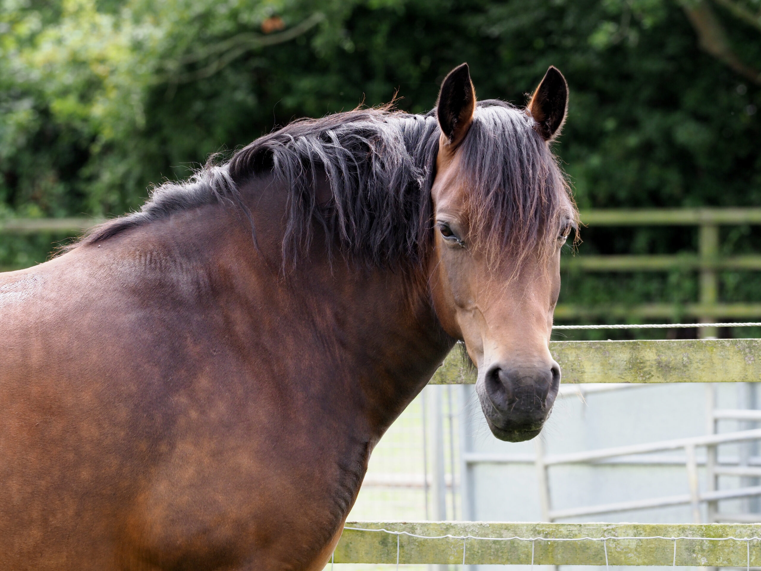
A common feature that puts horses at risk that owners can detect and address themselves before a laminitic crisis occurs is adipose deposited in certain areas of the horse’s body called regional adiposity. Regional adiposity describes fat or adipose tissue that is deposited in different regions of horses that owners should watch for if their horse is gaining weight. These common areas are the neck, commonly referred to as cresty necks, around the tail head, and sheaths of geldings or stallions. If these areas are noticed to be enlarging, especially in the spring when there is an abundance of fresh grass to graze on plus weight gain, then steps need to be taken to prevent the development of EMS.
Managing an Easy Keeper
It is very common to hear owners and veterinarians refer to heavy horses as easy keepers, but there can be some serious consequences of ignoring or brushing it off as just an easy keeper horse. Simple steps can be taken to reverse or reduce the risk of horses developing EMS by decreasing daily caloric intake. First, it is recommended to remove all grain from a horse’s diet including treats. Drastically reducing turn out time to graze especially fast growing lush grass is absolutely necessary in horses at risk of grass founder caused by EMS. There is some conflicting evidence as to when is the best time of day to allow a horse to graze that is sensitive to high sugars in lush growing grass. Some research has found that sugar levels peek later in the afternoon because of an abundance of sunshine and fully ramped up photosynthesis process that occurs in the grass. It is suggested then if grazing is allowed or deemed safe, that morning grazing is a safer time, but sometimes letting an at-risk horse graze is not worth the consequences. Other methods of allowing safe grazing are to mow the grass very short to minimize the volume of grass intake in a given period of time. If mowing is not an option, specially designed grazing muzzles allow pasture turn out but restrict the amount of grass taken in through the muzzle. Do not worry, as many horses are very quick to figure out how to get grass through the small hole in the muzzle and also allow the intake of water. It is recommended to have a leather poll strap on their halters to prevent injury when turned out while wearing a halter. Dry lot management is sometimes the only option, especially in horses that already have EMS. Keeping a horse on dry lot with no access to fresh grass and feeding more mature hay is sometimes needed to manage more severe cases. There are no current medications to help reduced the effects of insulin resistance due to obesity in horses, but some medications can be used to help with weight loss such as Thryo-L (levothyroxine) combined with consistent exercise.
Laminitis
Laminitis is the most debilitating and painful outcome of EMS, not to mention life threating. It is also the most expensive and difficult aspect of managing a horse with EMS. In order to properly manage laminitis caused by EMS, the horse needs to be examined by a veterinarian, radiographs need to be taken of the feet to assess the severity of the laminitis and an experienced farrier needs to be heavily involved with the management of the feet. If the disease is caught early, proper trimming may be all that is needed plus the other management aspects employed, of course, but in many cases corrective therapeutic shoeing is required. Pain management is another key aspect of addressing laminitis. NSAIDs or non-steroidal anti-inflammatory drugs, opioids, aspirin and an anticonvulsant drug called Gabapentin can help block or reduce pain of laminitis that horses experience. Some of these drugs do carry a risk of serious side effects so careful monitoring and proper dosages need to be on the order of a veterinarian to minimize the risk of side effects.
It cannot be repeated enough that the best cure for disease is through prevention. Taking early appropriate steps to keep horses from developing EMS is by far the best way to prevent the disease from occurring. If there is concern your horse is at risk of EMS, please talk to your veterinarian to determine if management and diet changes need to be made to prevent the development of this disease.
By Summer McMillen
As the land starts to thaw and cowboys and cowkids everywhere are gearing up for spring there is one specimen in particular that is dreading the coming season. And that is ponies. Or more specifically, kid horses.
Let’s look at life from the kid horses point of view for a second.they have the winter off. They’ve gotten to enjoy some much needed R&R in the back pasture. Their hair has gotten long and so have their hooves. They’ve spent the winter feasting on native grasses, alfalfa, and the occasional bucket of grain when it’s was especially cold.
They’ve gotten to wonder aimlessly while the real work horses were still being caught for essential tasks. The most work these kid horses have had to endure the last few months were being tied to the arena fence to get the cockleburs brushed out of their tail.
Yes, life has been calm for these kid horses. But, there is a season for everything and the season for being used and abused is fast approaching.
My own children all share a little pony they affectionately call “Rubble.” He is a certified welsh pony that is as tall as he is wide and is as passive as he is…not. They spend summers riding him bareback. And when they aren’t riding him bareback they are riding him fully saddled. Tiny little bit in his mouth being yanked in every which direction.
The little fellow takes everything in stride. But I can feel him starting to resent me from the back pasture already. Every time I carry him some feed he gives me a look that just screams “please keep all three of your children away from me forever please.” I laugh at him and tell him he doesn’t mean it and then he runs away as fast as he can. It’s a special bond.
Kid horses are funny like that. We entrust them with the lives of those most special to us. We saddle them up and throw the little cowkids on them and assign them small tasks like moving 200 pairs to a new pasture or, holding the herd while dad doctors wheat cattle. More often than not, the young partners get their job done. Sometimes in unlikely ways.
Last summer my daughter hopped up on Rubble bareback and was going to trot down the road and get our mail. A simple and harmless task I thought. I was proud of her for taking the initiative. I was also proud of her when I looked out the window, saw the little pony start crow hopping with excitement and the unassuming 8-year-old being catapulted in the ditch. Instead of crying she climbed back on. Tenacity, I thought. That’s what kid horses are good at teaching.
A few months later I was watering all our geldings. The three kids whom I raised, better known as the three amigos, were all harassing the little pony. My youngest son decided it was his turn. He climbed up on Rubbles back as proud as a peacock. But, he couldn’t help himself. He wanted to look that pony in the eye. So he starts leaning. Farther and farther until he leaned all the way to the ground. Lost a boot in the process. Instead of rushing to his side I waited for the tears. He was only two years old at the time after all. Instead his face was filled with glee. Joy, I thought. That’s what kid horses are good at teaching.
My middle boy is afraid of heights. So mounting a horse has been tough for him. When we introduced Rubble, I wasn’t sure he could even manage his height. After a little encouragement he donned his chaps, boots, and hat with a turkey feather in it and mounted the little horse. In a matter of five minutes the little cowboy had rubble trotting around the arena with his hand in the air and his spurs in his neck. (Gently of course.) Confidence builder, I thought. That’s what kid horses are good at teaching.
While, most of the time kid horses are a nuisance. Eating all our alfalfa and requiring special horse shoes to function properly.. they are more often than not a priceless gift. Teaching the ones we love most how to have tenacity, joy, and confidence.
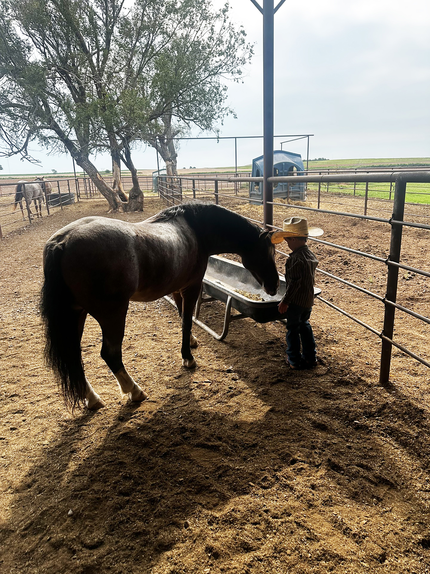
While the ponies in our life may love their time off in the winter you can’t convince me that they don’t love their jobs in the spring. Even if it’s just a little bit. When that little cowboy shoves a bucket of grain in their face after a job well done together, the ponies head sits a little higher and so does that little cowboys hat.

By Garrett Metcalf, DVM
A foot abscess is a common occurrence in horses throughout the year, with wet weather often contributing to an increase in cases. These abscesses can cause significant pain, lameness, swelling, and overall misery, making it important to address them quickly and manage pain to keep the horse comfortable. There are various methods used to treat foot abscesses, and this article will outline techniques to evaluate and treat them as efficiently as possible.
A foot abscess is a localized or sometimes diffuse infection trapped between the sensitive and non-sensitive laminae within the hoof capsule. Abscesses may develop spontaneously due to everyday stress and environmental factors that allow bacteria to penetrate down to the sensitive tissues. Other causes include penetrating injuries to the sole from nails, sharp rocks, or even thorns. Poor hoof care and misdriven shoeing nails can also lead to abscess formation. Common sites include the white line, where the sole and hoof wall meet, and the bars of the heels.
The level of lameness caused by a foot abscess can vary, but it often results in visible discomfort at the walk and can even cause non-weight-bearing lameness. Swelling that begins at the foot and moves up the leg may occur, particularly if the abscess migrates and ruptures at the coronary band. These cases are often referred to as “gravel” abscesses, which are simply abscesses that find the path of least resistance and exit at the coronary band, creating a draining tract. In some cases, especially involving a hind foot, the horse’s movement may appear so abnormal that it mimics neurological issues, confusing owners and veterinarians.
Diagnosing a foot abscess begins with a lameness exam. Most affected horses will be visibly lame at the walk, though in some cases a trot may be necessary to detect the issue. Regional nerve blocks can help confirm that the pain is originating from the foot and not another part of the limb. Horses with abscesses often show an increased digital pulse and, occasionally, noticeable heat in the foot. The bounding pulse is due to inflammation and is most easily felt just above the hoof near the ankle. If the horse is shod, removing the shoe is often necessary for a thorough exam. Hoof testers are useful in identifying the most painful area, and horses with abscesses are typically reactive to pressure. Cleaning out the foot with a hoof knife is important for exposing any defects or tracts in the sole or frog. Often, a dark spot or line will lead to the source of the abscess.
There are multiple ways to treat an abscess, and opinions vary widely, but my preferred approach is to open the abscess as soon as possible. This provides nearly immediate relief for the horse and allows the infection to begin resolving. A sharp hoof knife or loop knife is a reliable tool to open the abscess and create drainage through the bottom of the foot. Allowing the abscess to drain from the sole reduces the risk of a gravel abscess and makes it easier to treat the area with topical poultices. After opening, it’s important to bandage the foot both to draw out remaining infection and to keep the area clean.
A large baby diaper makes a simple, effective bandage. It’s absorbent and fits the hoof well. Secure it with layers of Vetrap, duct tape, and Elastikon, or place the hoof in a medicine boot to keep it protected. Poultice choice is often based on personal experience and availability, but the goal is the same — to draw out infection and prevent contamination. Products like Magna Paste, an Epsom salt-based poultice, are effective, as is a homemade mix of sugar and Betadine. There are many other options, but whatever product is chosen should be safe and offer antimicrobial properties.
In some cases, an abscess may be difficult to locate or open. Soaking the foot in warm Epsom salt water can help soften the hoof and encourage the abscess to rupture or become easier to identify. Pain management is also helpful while waiting for the abscess to surface. If the abscess continues to recur or proves difficult to treat, radiographs can help evaluate the hoof’s internal structures. While most abscesses don’t show up on X-rays — since the fluid is the same density as the hoof — they may be visible if gas is present within the abscess. Radiographs are especially important in cases of puncture wounds, to ensure deeper structures like the coffin joint or navicular bursa aren’t involved. In cases of penetration, it’s best to leave the foreign object in place until X-rays are taken, which helps determine the extent of the injury and what structures may have been affected.
While preventing all foot abscesses isn’t always possible, good hoof care goes a long way. Regular trimming on a consistent schedule helps maintain healthy laminae and prevents stretching of the white line, which can allow bacteria to enter. Careful shoeing practices, including proper nail placement, can further reduce the risk of abscess development.
Foot abscesses are painful, frustrating, and often sudden — but with proper diagnosis, drainage, and aftercare, horses typically recover well and quickly return to soundness.
When I was younger, I saw plenty of old Westerns. They were fun to watch, but one part always stressed me out. Inevitably, a cowboy would get shot or thrown from his horse, and while the cameras stayed on the fallen rider, I worried about the horse. Would it wander around lost on the prairie, never finding its way back? The truth is, most horses know exactly where home is. Turn one loose, and it will drift toward the barn. Ride one out, and the trip away from home feels steady, but the return picks up pace the moment the barn roof comes into sight. We even have a name for it: barn sour.
Horses are prey animals, and survival has always depended on familiar ground. For a domestic horse, the barn means food, water, and the company of the herd. Ethologists (scientists who study animal behavior) point out that horses are quick to learn patterns. When hay and grain appear in the same place every day, that spot becomes magnetic. Over time, repetition lays down mental trails as clearly as cattle wear down physical ones in a pasture. What appears to be stubbornness is actually instinct. The barn equals safety, and safety equals survival. Riders from cavalry days to modern ranches have written about horses quickening their pace on the way home. And though the land changes, that pull never does.
People are not so different. We all have barns in our lives — comfort zones we gravitate toward, routines that steady us. They serve a purpose. Like a horse standing at the gate, we lean on safe ground when life feels uncertain. But the pull can also hold us back. A horse that refuses to leave the yard never discovers what lies beyond the fence, and the same is true for us.
That balance shows up in history too. Old cattle trails once served their purpose, guiding herds north and helping to build economies. But when railroads and fences changed the landscape, those well-worn tracks became ruts. Progress required new paths. Our own habits work the same way. Some keep us grounded. Others only circle us back to where we started.
When I see my horses drifting toward the barn, I think less about impatience and more about instinct. They are drawn to the familiar, and so am I. The barn matters. It is the anchor point, the place of rest. But the pasture matters too, because growth is waiting outside the gate.
Those old Westerns had it right in at least one way. The cowboy’s horse was never going to wander off aimlessly. It would head back to camp, back to the barn. That simple truth still plays out in every pasture and arena today. Horses know where home is. The question is whether we will let the pull of our own barns keep us tied too tightly, or whether we will use them as a base to step farther into the wide-open ground ahead.


-
 Country Lifestyle2 years ago
July 2017 Profile: J.W. Hart
-
 Attractions9 years ago
48 Hours in Atoka Remembered
-
 Equine9 years ago
Umbilical Hernia
-
 Outdoors8 years ago
Grazing Oklahoma: Honey Locust
-
 Country Lifestyle4 years ago
The Two Sides of Colten Jesse
-
 Farm & Ranch7 years ago
Hackberry (Celtis spp.)
-
 Farm & Ranch1 year ago
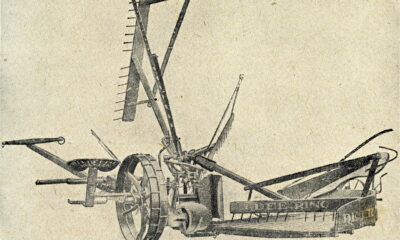
From Plow to Plentiful: The Most Important Inventions in Agricultural History
-
 Country Lifestyle10 years ago
The House a Treasure Built