
Farm & Ranch
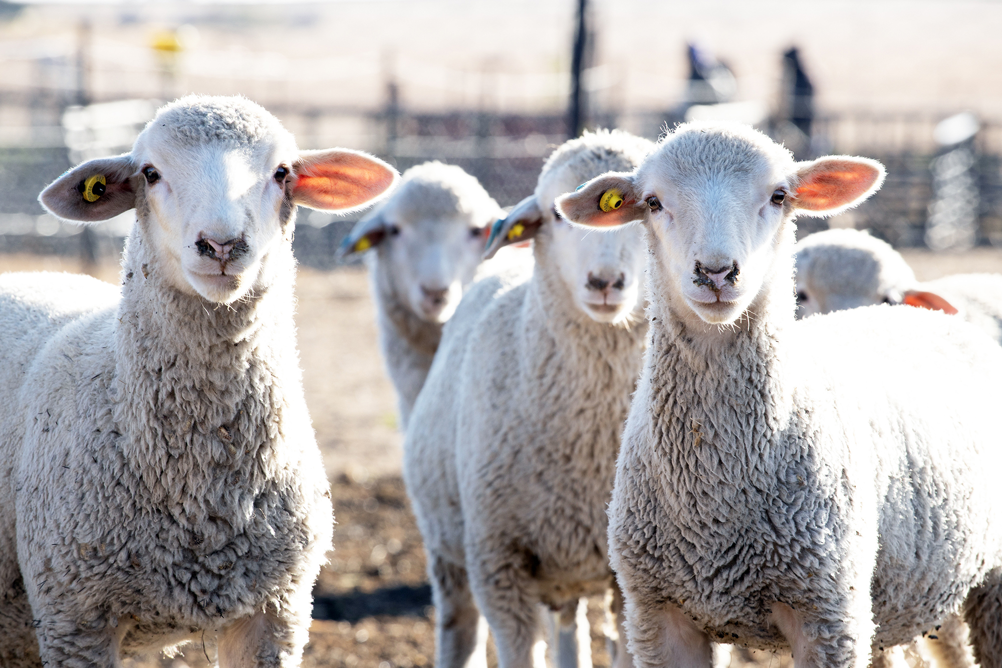
Footrot in Sheep and Goats

Barry Whitworth, DVM, MPH | Senior Extension Specialist | Department of Animal & Food Sciences | Ferguson College of Agriculture | Oklahoma State University
Foot issues are a common finding in sheep and goat operations. According to a 2011 Sheep Study conducted by the United States Department of Agriculture, footrot was found in 37.3% sheep operations. In the 2009 Goat Study, “symptoms of sores on hoof area with foul odor” were found in 6.6% of all the operations. Some studies indicate the issue increases with herd size.
Several factors contribute to the disease, but the main bacterium involved is Dichelobacter nodosus. The bacterium is contagious and does not survive long outside the host. However, the bacterium will live for years in the feet of sheep and goats. The bacterium has many strains. In general, they can be classified as benign or virulent. Virulent strains are able to destroy the horn. Benign strains do little or no damage to the horn.
Two other bacteria may play a role in footrot. Fusobacterium necrophorum, which causes foot scald, is a common inhabitant of the digestive tract of ruminants. It does not appear to be contagious. This bacterium usually contributes to the development of footrot. Some experts believe that foot scald is a precursor to footrot. One other bacterium that may contribute to the development of footrot or footscald is Trueperella pyogenes. This bacterium may increase the susceptibility of the hoof to the other two bacteria. This bacterium is found in the environment and is associated with foot abscesses.
The first sign of foot scald is usually lameness. In foot scald, the space between the claws may appear red, hairless, swollen, and moist. No odor is present, and the condition tends to improve with dry weather.
In comparison, footrot will have the same clinical signs as foot scald but the signs will be more severe. The space between the claws will be necrotic and the hoof will be affected. The foot will have a foul odor. In severe infection, the hoof wall will separate from the pedal bone. The condition is highly contagious and will spread rapidly through the flock.
Both footrot and foot scald are usually diagnosed on clinical signs. The presence of a foul odor is important to separate footrot from foot scald. A definitive diagnosis is based on culturing the bacteria from the foot.
Several management tools need to be used when dealing with footrot. Studies show a significant improvement in overall herd lameness when lame animals were separated from the flock. Sheep infected with Dichelobacter nodosus that causes footrot continue to shed the organism for a period of time after treatment. If left in the herd, these animals contaminate pastures and facilities which contribute to the spread of bacteria. Lame animals need to be isolated until they are healed.
Research has demonstrated that there is a benefit to early identification and prompt treatment of lame sheep. Sheep need to be observed daily for health issues. Studies show that treatment should be initiated within 3 days of lameness. Any animal found to be lame needs to be treated promptly with a long-acting antibiotic and topical antibiotic. Their feet should not be trimmed at this time. Trimming infected feet delays healing.
Another management tool is the use of foot baths. Using a foot bath during a footrot outbreak has been found to reduce lameness. Copper sulfate, formalin, and zinc sulfate may be used in foot baths, but a 10% zinc sulfate solution is preferred.
Lastly, significant improvement in a flock can be made by culling lame sheep. Sheep requiring 2 or more treatments should be culled. Removing chronically infected sheep from the flock reduces a significant source of the bacteria which should lower the spread of the disease. Culling will improve the economics of the flock by decreasing labor time and money associated with treating footrot. It is also recommended not to keep lambs from culled ewes with footrot for replacement ewes.
Preventing footrot begins with proper nutrition. Hoof health is influenced by minerals and vitamins. Special emphasis should be placed on ensuring adequate amounts of selenium, biotin, zinc, Vitamin A, and Vitamin E. Hooves should not be allowed to over grow. Routine trimming and examination will ensure a healthy foot. Facility and pasture management play an important role in controlling lameness as well. It was found in a study conducted in the United Kingdom that allowing pastures to rest 2 or more weeks had a high impact in reducing lameness in sheep. Facilities need to be kept clean and dry. Lambing pens need to be cleaned and disinfected between uses. Fences need to be maintained. This prevents neighboring animals from entering the farm which could contaminate the pasture with unwanted bacteria.
Footrot can be a frustrating disease. However, following the above practices should improve foot health. For more information about footrot in sheep and goat enterprises, producers should consult with their veterinarian or the Oklahoma State University County Cooperative Extension Service Agriculture Educator.
References
Reilly, L. A., Baird, A. N., & Pugh D., G. (2002). Diseases of the foot. In D.G. Pugh(Ed.), Sheep & Goat Medicine (pp. 225-227). W.B. Saunders Company
Witt J, Green L. Development and assessment of management practices in a flock-specific lameness control plan: A stepped-wedge trial on 44 English sheep flocks. Prev Vet Med. 2018;157:125-133.
By OKFR Staff
Livestock One of the most serious late- summer concerns is a bloom of cyanobacteria, commonly called blue-green algae. These organisms are naturally present in many bodies of water, and most do not produce dangerous toxins. The risk increases when certain toxin-producing species multiply rapidly under favorable
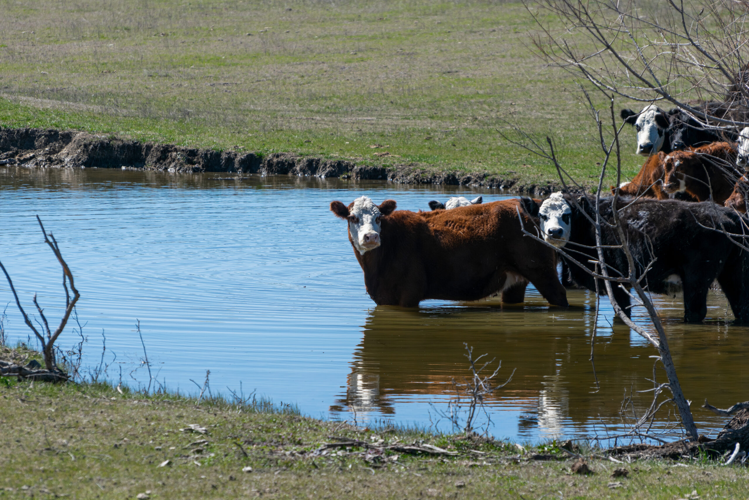
During the hottest part of summer, a farm pond can look like a welcome source of water. Cattle may be standing belly- deep along the bank, and the pond may still hold enough water to appear dependable. However, appearance alone does not reveal whether that water is safe.
Hot weather, limited rainfall and falling pond levels can create water-quality problems. As water evaporates, salts, minerals, nutrients and other substances may become more concentrated. Warm, still water can also encourage the growth of blue-green algae, some forms of which can produce toxins capable of killing livestock.
Water is sometimes treated as an afterthought compared to grass, hay and supplemental feed. In reality, livestock need a dependable supply of clean water to maintain feed intake, regulate body temperature and support production. Poor-quality water may reduce consumption and performance even when it does not cause an obvious case of poisoning.
A pond that has watered cattle for years should not automatically be considered safe under every set of conditions. Producers should pay particular attention during drought, extreme heat or any period when the water level drops significantly.
Watch the Water and the conditions.
According to Oklahoma State University Extension, toxic blue- green algal blooms typically develop in standing water following periods of hot, dry and calm weather. Nutrients such as nitrogen and phosphorus can contribute to excessive growth. Those nutrients may enter a pond through manure, fertilizer runoff, soil erosion and decaying vegetation.
A suspicious bloom may make the water appear bright green, blue-green, pea-soup green or even brownish. Scum, foam or mats may collect along the shoreline, particularly where the wind pushes floating material into one area. Some blooms resemble spilled paint.
Not every green pond is poisonous. Common aquatic plants and harmless forms of algae can also change the color or surface of the water. Unfortunately, it is usually impossible to determine whether a bloom is toxic simply by looking at it. A pond can contain toxin-producing cyanobacteria without displaying every commonly described warning sign.
Blue-green algae can produce toxins that affect the nervous system or liver. Depending on the toxin and the amount consumed, affected animals may develop weakness, staggering, difficulty breathing, tremors, convulsions, diarrhea or collapse. In severe cases, animals may be found dead near the water before the owner realizes anything is wrong. Treatment is often unsuccessful, making prevention especially important.
Any suspected poisoning should be treated as an emergency. Remove the herd from the water source without driving or unnecessarily stressing animals that may already be affected. Provide another clean water supply and contact a veterinarian immediately. The local county Extension office can also help determine where and how to submit an appropriate water sample.
Blue-green algae is not the only potential problem. Pond water may contain elevated levels of dissolved salts, sulfates, nitrates, bacteria or other contaminants. Runoff can carry manure, fertilizer, pesticides and organic matter into the pond. Low water levels can make some of these problems more pronounced because the remaining water becomes increasingly concentrated.
Water with excessive dissolved solids may taste bitter or salty, reducing the amount animals consume. Depending on the substances involved and their concentrations, poor- quality water may contribute to diarrhea, reduced feed intake, poor weight gain or other health concerns. Young animals and livestock that are not accustomed to the water may be more vulnerable.
Producers should also watch the cattle themselves. Are they reluctant to drink? Are they crowding around a different water source? Has feed intake or weight gain declined without an obvious explanation? Are several animals experiencing digestive problems? Changes involving multiple animals can provide an early clue that feed or water should be investigated.
OSU Extension recommends collecting a representative sample and submitting it for a livestock water test when a water supply is suspected of harming animal health or performance. A standard livestock water test may evaluate several characteristics, but blue-green algae concerns can require different sampling and testing procedures. Producers should contact the laboratory or county Extension office before collecting the sample so it is taken from the correct location, placed in the proper container and handled appropriately.
They Develop Good pond management cannot eliminate every water- quality problem, but it can lower the risk. One of the most effective steps is limiting the amount of manure, soil and nutrients that enter the water.
Allowing cattle unrestricted access to an entire pond can result in damaged banks, muddy water and large amounts of manure being deposited in or near the water. Hoof traffic removes vegetation and contributes to erosion. Sediment fills the pond over time, while manure supplies nutrients that may encourage excessive algae growth.
Where practical, fencing livestock away from most of the pond and providing a limited-access watering point can improve conditions. Another option is piping water to a trough located below the dam or elsewhere in the pasture. OSU notes that reducing cattle excrement in the pond can decrease conditions favoring toxic algae and improve water palatability.
A vegetated buffer around the pond can also slow runoff and help trap soil and nutrients before they reach the water. Fertilizer and manure should be applied carefully in the water
shed, particularly before heavy rainfall. Producers should avoid allowing large amounts of hay, feed or organic debris to accumulate at the water’s edge.
Alternative watering systems require maintenance, too. Troughs should be checked for algae, manure, drowned animals and other contamination. Wildlife escape ramps should be installed in tanks so birds and small animals can climb out rather than drowning and contaminating the water.
Owners should be cautious about treating a pond on their own. Copper sulfate and other algaecides are sometimes used for algae control, but improper treatment can create additional problems. Killing a large quantity of algae or aquatic vegetation at one time can reduce dissolved oxygen as the material decomposes, potentially leading to a fish kill. Treatment may also release toxins from affected cyanobacteria into the water. Any chemical application should be based on proper identification and professional recommendations rather than guesswork.
Livestock should always have access to enough water for the weather, their size and their stage of production. Demand increases sharply during hot weather, and a pond that barely meets the herd’s needs in spring may not be sufficient in August. Check the source regularly rather than waiting for cattle to gather around a muddy shoreline.
Looks can be deceiving when it comes to ponds. Crystal-clear water isn’t always safe, and a green pond doesn’t necessarily mean toxic algae.
That’s why regular observation, testing and proactive prevention are essential for maintaining pond.
By Barry Whitworth, DVM
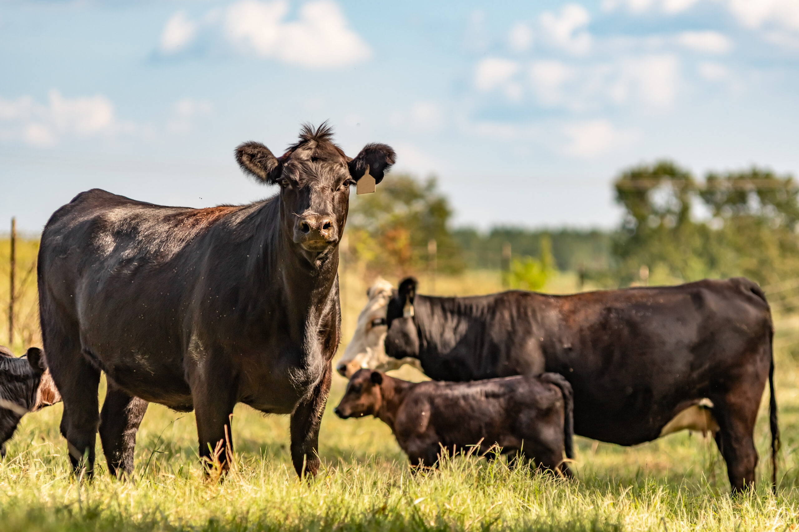
Over the past few years, Theileria orientalis has been detected in cattle in Oklahoma. Early cases were primarily diagnosed in adult cattle. However, more recent cases in Oklahoma have been identified in stocker cattle. All cattle producers should be familiar with the clinical signs of this disease and observe their cattle daily for any signs of trouble.
In the United States, the first case of theileriosis was identified in Virginia in August 2017. These cattle exhibited clinical signs of weakness and anemia. An initial diagnosis of anaplasmosis was made. Blood samples from the animals were tested for Anaplasma, Babesia, and Leptospira. Test results were negative for all three; however, a blood protozoan was detected. This organism was identified as T. orientalis genotype Ikeda. Since this initial herd outbreak, the organism has been detected in several states.
Cattle infected with and showing illness from T. orientalis genotype Ikeda may exhibit clinical signs such as fever, weakness, anorexia, and exercise intolerance. If forced to move, affected cattle may stagger and gasp for air. If stressed excessively, they may collapse and die. Upon examination, the gums, eyes, or vaginal mucosa may appear pale (white) or yellow in color. Reproductive losses, including stillbirths and late-term abortions, may occur, along with a reduction in milk production.
Clinical signs reported in adult cattle in Oklahoma include anemia, sudden death, weight loss, abortion, failure to thrive, and failure to calve. In stocker cattle, weight loss, anemia, poor performance, and sudden death have been reported.
Because Anaplasma marginale and T. orientalis genotype Ikeda produce similar clinical signs, distinguishing between the two can be challenging.
One notable difference is that clinical signs are more commonly observed in young cattle infected with T. orientalis, whereas this is less common in cattle infected with A. marginale. Additionally, cattle with anaplasmosis often display aggression, whereas those with T. orientalis genotype Ikeda typically do not. A laboratory test is required to definitively differentiate between the two diseases.
Haemaphysalis longicornis has been identified as a possible vector of T. orientalis genotype Ikeda. In 2017, the United States Department of Agriculture’s (USDA) National Veterinary Services Laboratories (NSVL) confirmed the presence of H. longicornis, commonly referred to as the Asian longhorned tick (ALT) or bush tick. In efforts to determine how the tick arrived in the United States, USDA officials discovered it had been present in West Virginia as early as 2010. The tick has now been confirmed in at least 26 states, including Oklahoma. There is also some evidence suggesting additional insect vectors may transmit T. orientalis genotype Ikeda. Needle transfer is another possible route of transmission.
The ALT has been identified in Craig and Mayes counties in Oklahoma. This tick thrives in areas with high humidity, such as wooded regions, brush, or tall grass. Ticks are often found where large numbers of wildlife congregate, such as along deer trails. Producers seeking more information about the ALT can visit the USDA website at: https://www.aphis.usda. gov/livestock-poultry-disease/ cattle/ticks/asian-longhorned.
In other countries, treatments have been developed for T. orientalis. Unfortunately, no approved treatments are currently available in the United States, and no vaccines exist for this disease. The best defense is prevention: purchasing cattle free of the organism, avoiding blood transfer between animals, and implementing effective tick control measures. Producers should purchase cattle from reputable sources. Instruments should be cleaned between animals during procedures such as castration or dehorning, and needles should always be changed between animals. External parasites must be controlled using appropriate insecticide treatments. In addition to insecticides, pasture management strategies—such as rotation to avoid wooded or brushy areas where ticks thrive—are important. Patch burning may also help reduce tick populations.
Theileria orientalis remains a concern for some cow/calf and stocker producers in Oklahoma. Producers seeking more information about T. orientalis genotype Ikeda should contact their local veterinarian or Oklahoma State University County Extension Agriculture educator.
References Hammer, J. F., Emery, D., Bogema, D. R., & Jenkins, C. (2015). Detection of Theileria orientalis genotypes in Haemaphysalis longicornis Ticks from Southern Australia. Parasites & vectors, 8, 229.
Oakes, V. J., Yabsley, M. J., Schwartz, D., LeRoith, T., Bissett, C., Broaddus, C., Schlater, J. L., Todd, S. M., Boes, K. M., Brookhart, M., & Lahmers, K. K. (2019). Theileria orientalis Ikeda Genotype in Cattle, Virginia, USA. Emerging infectious diseases, 25(9), 1653–1659.
Spickler, Anna Rovid. (2019). Theileriosis. Retrieved from http://www.iastate.edu/DiseaseUbfi/factsheets.php.
Watts, J. G., Playford, M. C., Hickey, K.L. (2016). Theileria orientalis: A Review, New
Zealand Veterinary Journal, 64:1, 3-9.
By Ann Asher
Then there are the diseases and pests that younger generations have only read about.
For decades, New World screwworm belonged in that second category. Older ranchers remembered the days when a simple branding wound or a newborn calf’s navel had to be watched closely because it could attract one of the livestock industry’s most destructive pests. Veterinarians still taught students how to recognize it, but for many producers, screwworm was something that had been conquered long before they ever worked their first set of cattle.
This summer, that changed. On June 3, 2026, the U.S. Department of Agriculture confirmed New World screwworm in a calf in South Texas, marking the parasite’s return to the United States after decades of successful eradication. While Oklahoma has not reported any confirmed cases, the discovery has ranchers across the Southern Plains paying close attention. The concern isn’t just where screwworm is today, but where it could be tomorrow.
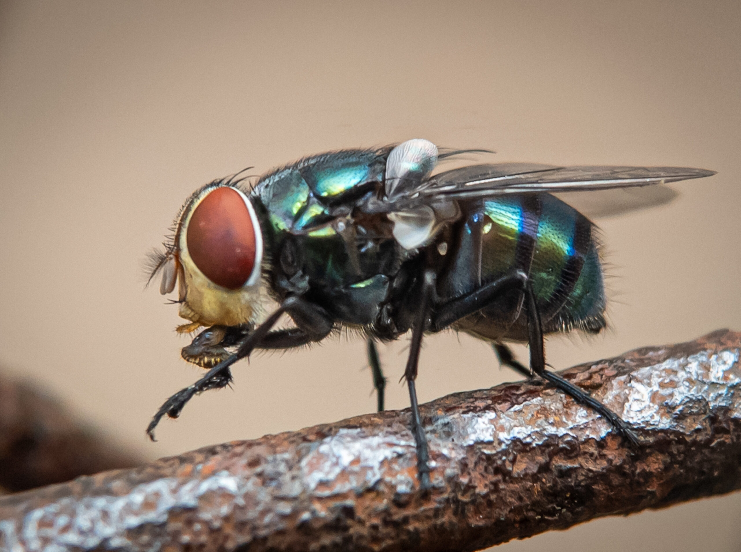
Unlike the common maggots that feed on dead tissue, New World screwworm larvae feed on living flesh. Adult female flies are drawn to fresh wounds and natural body openings, laying hundreds of eggs at a time. Within hours, the larvae hatch and begin burrowing into healthy tissue, enlarging the wound as they feed. Left untreated, an infestation can become life-threatening.
There are some livestock problems that today’s ranchers know by heart. Pinkeye. Foot rot. Horn flies. Internal parasites. Most producers have dealt with them at one time or another.
The list of potential hosts is long. Cattle, horses, sheep, goats, pigs, dogs, wildlife and, in rare cases, people can all become infested. Even relatively small injuries—a wire cut, a tick bite, a branding wound, a castration site, a dehorning wound or a newborn calf’s healing navel—can provide an opportunity for the fly.
Fortunately, today’s producers have one advantage their grandparents often did not: they know what they’re looking for.
A wound that continues to grow instead of heal should never be ignored. Bloody or watery discharge, a strong foul odor, swelling, pain, visible larvae and unusual behavior such as excessive licking, head shaking or isolation from the herd all warrant a closer look. While these signs can indicate several different conditions, they should also prompt producers to consider the possibility of screwworm and contact their veterinarian immediately.
The return of screwworm has also reminded many people of one of agriculture’s greatest success stories.
During the mid-1900s, New World screwworm cost American livestock producers millions of dollars each year. Rather than relying solely on insecticides, scientists developed what became known as the Sterile Insect Technique. Millions of sterile male flies were raised and released into affected areas. Female screwworm flies mate only once during their lifetime. When they mated with sterile males, no offspring were produced. As generation after generation failed to reproduce, screwworm populations steadily declined until the pest was eliminated from the United States and later pushed farther south through Mexico and much of Central America.
That same strategy is once again leading the response.
Federal and state animal health officials have increased surveillance in affected areas, established movement controls where necessary and resumed sterile fly releases to prevent the pest from becoming established. Veterinarians, livestock markets and producers are all part of the early detection system, with every suspicious case helping officials determine where additional monitoring may be needed.
For Oklahoma producers, the message is one of awareness rather than alarm.
As of this writing, no cases have been confirmed in Oklahoma. Even so, Oklahoma shares a long border with Texas, and livestock routinely move throughout the region for sales, breeding, shows and grazing. That makes vigilance especially important during fly season.
Routine herd checks have always been one of the best management tools available, and they are even more valuable now. Fresh wounds should be treated promptly and monitored until they heal. Newly branded or processed cattle deserve extra attention, as do newborn calves, horses with injuries, sheep after shearing and any animal recovering from surgery or trauma. Wildlife can also serve as hosts, making complete prevention impossible, but early detection can make all the difference.
If producers suspect screwworm, they should avoid simply treating the wound and moving on. Instead, they should contact their veterinarian or the Oklahoma Department of Agriculture, Food and Forestry so the case can be evaluated and, if necessary, samples collected. Rapid reporting is one of the most effective tools available for limiting the spread of the pest.
Perhaps the most encouraging part of this story is that agriculture has faced this challenge before—and won.
The same science that eliminated New World screwworm from the United States decades ago is once again being deployed. Researchers understand the insect’s life cycle. Veterinarians know what to look for. Producers are better connected than ever through Extension services and state animal health agencies.
Screwworm may have returned to the United States, but so have the tools that defeated it the first time.
For Oklahoma livestock producers, the best response is also the simplest: keep an eye on your animals, don’t ignore wounds that aren’t healing, and report anything that doesn’t look right. When it comes to screwworm, finding one case early is far better than finding many too late.





-
 Country Lifestyle2 years ago
July 2017 Profile: J.W. Hart
-

 Farm & Ranch2 years ago
From Plow to Plentiful: The Most Important Inventions in Agricultural History
-
 Attractions9 years ago
48 Hours in Atoka Remembered
-
 Equine9 years ago
Umbilical Hernia
-
 Country Lifestyle5 years ago
The Two Sides of Colten Jesse
-
 Outdoors9 years ago
Grazing Oklahoma: Honey Locust
-
 Farm & Ranch8 years ago
Hackberry (Celtis spp.)
-
 Equine6 years ago
On the Road with Emily Miller-Beisel