Equine
Living Her Dream
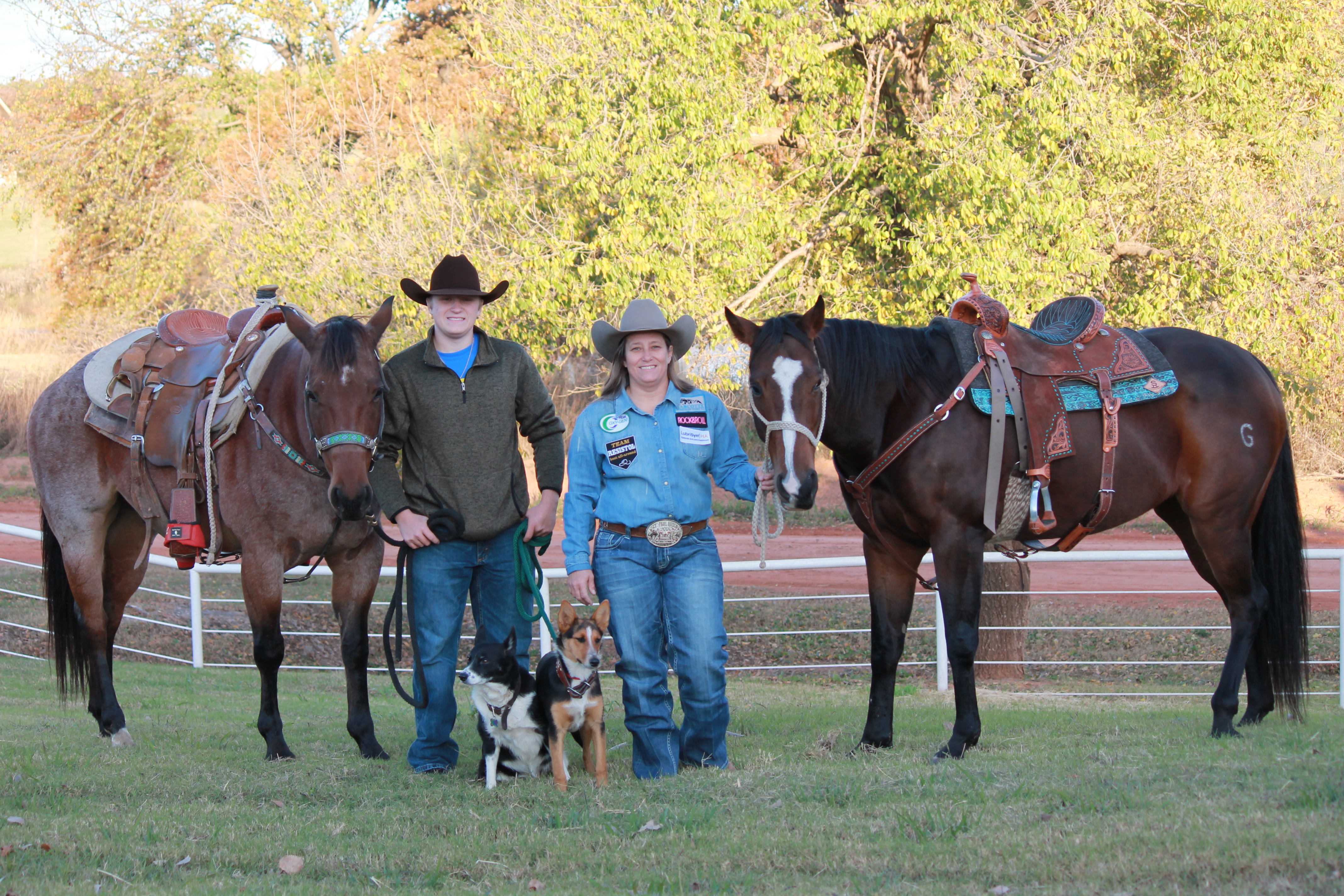
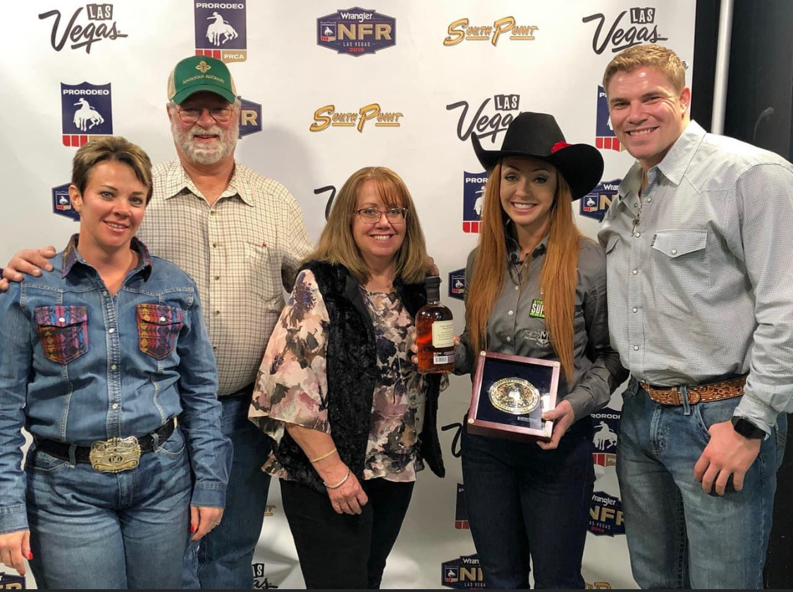
On the eve of her first Wrangler National Finals Rodeo appearance, Tracy Nowlin feels relief. The accomplishment, although major, was tough to earn and a long time coming. She won numerous large rodeos earning over $90,000 in fewer than 80 events, finishing twelfth in the World Standings. More impressive is that she did it all on one very special horse.
A Rodeo Upbringing
Nowlin grew up as part of one of Oklahoma’s most familiar rodeo families. Her father, Terry Postrach Sr., was a well-known calf roper, and her brother, Terry Postrach Jr., was a three-time International Professional Rodeo Association World Champion calf roper. Her mother, although not a competitor herself, might as well have a gold buckle for all the cowboys and cowgirls she helped haul to world championships. Tracy’s son Ty, an 18-year-old high school senior, is an accomplished calf roper.
Tracy herself has made the International Finals Rodeo in Oklahoma City 14 times, closing in on the record of 16 held by fellow Oklahoma barrel racer Betty Roper.
It’s not surprising Nowlin grew up to be a cowgirl, although her unwavering love of rodeo might be. “Growing up we roped until midnight and never knew anything else. I never was a normal kid. I got up in the morning and saddled my horse. They never had to tell me to ride, but they’d have to tell me that was enough and that it was time to do school work or go to bed,” she recalled.
With two ropers in the family, Nowlin spent many hours working chutes and traveling to ropings. “We roped until midnight and never knew anything else. There are eight years between my brother and me, and he was my best friend until he went off to college. We did everything together,” she explained.
Although roping was a major part of her life, the young cowgirl managed to qualify for her first IFR at only 13 years old with a horse named Goose. She qualified for the finals three more times on Goose, who made his final trip to OKC at over 20 years old.
She went to the IFR three times on a gelding named Rambo, who did double-duty as her brother Terry’s tie-down horse. Her other mount was a mare named Dusty Rose, who went to the IFR six times.
Nowlin’s life could have taken a vastly different course. After high school, Tracy received a scholarship for art school. While tempting, the desire to rodeo was stronger. “I had a really nice barrel horse when I graduated, and I could have gone to Santa Fe for art school. I did Indian art, and I really wanted to do sculpture, but I didn’t. I regret not going some, but my first love was rodeoing. It always has been. I never wanted to do anything but be on a horse. My first real word was horse,” Nowlin explained.
They mother of a friend of hers taught her how to peyote, a bead-weaving technique. “I’ve beaded all my life. That’s how I survived; by making stuff when I didn’t have a barrel horse,” she said. Nowlin, a professional beader, still keeps busy in down time by beading for tack and other accessories. Although a retailer does sell her creations, many people buy directly from her.
A Special Horse
It was during one of those barrel horse-less times four years ago that a non-descript bay mare came into her life. At the time, Nowlin was looking for a new tie-down horse for her son Ty and found an 8-year-old mare on a Facebook Buy or Sale group. She was owned by a man named Shawn Howell from Welch, Okla., who was a fiddle player in the Texas Playboys.
The ad noted that the mare, listed for sale at $1,850, had been roped off of and had seen the barrels, so she looked like a possible match. “I was scared he was going to sell her before we were able to pick her up. He didn’t have any calves to rope, so he let us take her home to try,” Nowlin recalled. “She’d been turned out for three or four years in the pasture and hadn’t been rode much.”
When they tried her on cattle, it was apparent that the little mare wouldn’t work. Knowing she had seen the barrels, Nowlin spent some time riding her that day, but didn’t feel like she’d fit. “I was ready to send her back, but that night I couldn’t sleep. I had that gnawing feeling like you get when you know you’re about to make a big mistake,” she said. “I told God that, if he’d just let me go to sleep, I’d give her another try in the morning.”
When she rode the mare the next day, she was impressed that she’d retained what she’d been taught the night before. “I was trying every way to talk myself out of buying her because I really liked her, and I needed a barrel horse, but the momma in me said Ty needed a calf horse more than I needed her. God had a different plan though,” Nowlin said.
The mare, registered as DJG Maddison, reminded Nowlin of another horse she’d owned. “My mom asked me why I wanted her so bad, and I told her that she’s the only horse I’ve been on that is as quick-footed as the roan horse I had.”
Nowlin bought “Dolly Jo” at the end of October in 2014. After approximately a month of riding, she decided to take the mare to a race. “It was right before Christmas. I was doing a lot of bead-working at the time making a living and had gotten all my Christmas orders out, and decided I’d go to the barrel race and exhibition her,” Nowlin said. Exhibitions are used to get barrel horses experience away from home. The runs, while timed, are not eligible to win money.
Nowlin had planned to exhibition the mare three times that night, but after the mare worked so well the first time, she decided that was enough. The next week, she decided to enter the actual class. “We won the 2D the next week, and she didn’t go half as fast as we’d been going. It was only four or five weeks later that she started winning the 1D,” Nowlin said.
That meteoric rise to the top continued. At their first rodeo, the pair was just one or two spots out of placing. One of the next rodeos was Duncan, Okla., and after seeing the contestant roster, Nowlin began to have some doubts. “I came back to the truck and mom asked what was wrong. I told her I was worried we might have stepped off a little deep this time, because everyone who was good was entered. She told me not to lose my faith now,” Nowlin said. She and Dolly Jo wound up placing in the rodeo, just hundredths of a second off the reigning World Champion.
“After that, I knew she was something special, but I thought she’d just be a little pen horse because of her size. I thought I could go run her at jackpots until I got another horse ready, but it’s not how it worked out,” Nowlin said.
Read more about Tracy and Dolly Jo in the December issue of Oklahoma Farm & Ranch.
By Dr. Devan England DVM
Does your horse have gastric ulcers? Gastric or stomach ulcers are frequently blamed for a variety of things including poor performance, acting ‘cinchy’, weight loss, not eating, poor coat condition, diarrhea and colic. However, gastric ulcers are not always the culprit and the only way to know for sure if your horse has gastric ulcers is to look at the stomach on camera, using an endoscope. Poor appetite and poor body condition are the mostly widely observed clinical signs with gastric ulcers, however, these are non-specific. If you think your horse might have gastric ulcers, the best place to start is to talk to your veterinarian and consider scheduling a gastroscopy. Gastroscopy requires the horse be held off feed for at least 16-18 hours and held off water for at least 6-8 hours. Fasting off feed and water is necessary to allow the veterinarian to see the whole stomach. If restricting feed or water is difficult in your management situation, many veterinarians will allow you to hospitalize your horse the night before gastroscopy for proper fasting.
Gastric ulcers are split into two types, classified by the location of the ulcer in the stomach. Squamous ulcers are ulcers that occur in the squamous or skin like portion of the stomach. This is the top part of the horse’s stomach, is closest to the esophagus, and has squamous tissue to protect this portion of the stomach from stomach acids. The other ulcer type are glandular ulcers. Glandular ulcers occur in the bottom portion of the stomach, which is closest to the small intestine. This portion of the stomach has glandular mucosa with cells responsible for producing stomach acids for digestion as well as cells that produce mucus and buffers to protect the lining from stomach acid. Gastroscopy is important not only for diagnosing whether ulcers are present but also determining the severity and the type of ulcer, because these two ulcer types require different treatments.
Squamous gastric ulcers are common in racehorses both in and out of training, with higher prevalence in racehorses under training. Prevalence in Thoroughbred racehorses in training has been reported to be up to 100% (Sykes 2015). Squamous ulcers are also prevalent in Western pleasure horses, Thoroughbred stallions on breeding farms, and Italian donkeys (Sykes 2015). Glandular gastric ulcer prevalence has not been as well described as squamous ulcers. Glandular ulcers are reported to be most common in Thoroughbred and Standardbred racehorses, Canadian showjumpers and polo ponies, and American Quarter Horses (Sykes 2015).
Risk factors for ulcers vary by ulcer type. Anti-inflammatories (Bute, Banamine) can increase the risk of glandular ulcers in some horses by affecting normal defense mechanisms but are not a high risk in most horses. Horses that display stereotypic behaviors, such as cribbing, have an increased risk of squamous ulcers. Grain fed before hay in non-exercising horses, feeding larger amounts of grain, and increased time between meals increases the risk of squamous ulcers. Increased time with high intensity exercise and housing in single pens is associated with increased risk of glandular ulcers. A straw only diet, lack of water access and lack of direct contact with other horses increases the general risk of gastric ulcers.
If your horse is diagnosed with ulcers, the mainstay of treatment is a buffered formulation of omeprazole (Gastrogard, Ulcergard). Over the counter Omeprazole and compounded Omeprazole are not effective because without buffering, the acidic stomach quickly breaks down the drug before absorption. Most horses with squamous ulcers will have healing of these ulcers after a 4-week course of Gastrogard or Ulcergard at treatment dose (whole tube for the average horse). Some horses may be healed by 3 weeks of treatment, but all horses should undergo a recheck gastroscopy before stopping treatment. Horses diagnosed with glandular ulcers need combination therapy with Gastrogard/Ulcergard and Sucralfate for 4 weeks. About 2/3 of horses with glandular ulcers will heal in this time, but some horses may require longer treatment times so a recheck is always recommended before discontinuing treatment.
Horses at higher risk of gastric ulcers may benefit from preventative (low) doses of Ulcergard (1/4 tube in average sized horse) given for a few days before and during high stress situations like long distance travel and competitions. Sea buckthorn berry supplement may be protective against formation of glandular ulcers. Dietary management to decrease the risk of ulcers includes providing more frequent small hay meals if pasture access is not available, limiting high sugar grains as much as possible and adding vegetable oil to the feed.
Sykes BW, Hewetson M, Hepburn RJ, Luthersson N, Tamzali Y. European college of equine internal medicine consensus statement – equine gastric ulcer syndrome in adult horses. J Vet Internal Med 2015; 29:1288-1299.
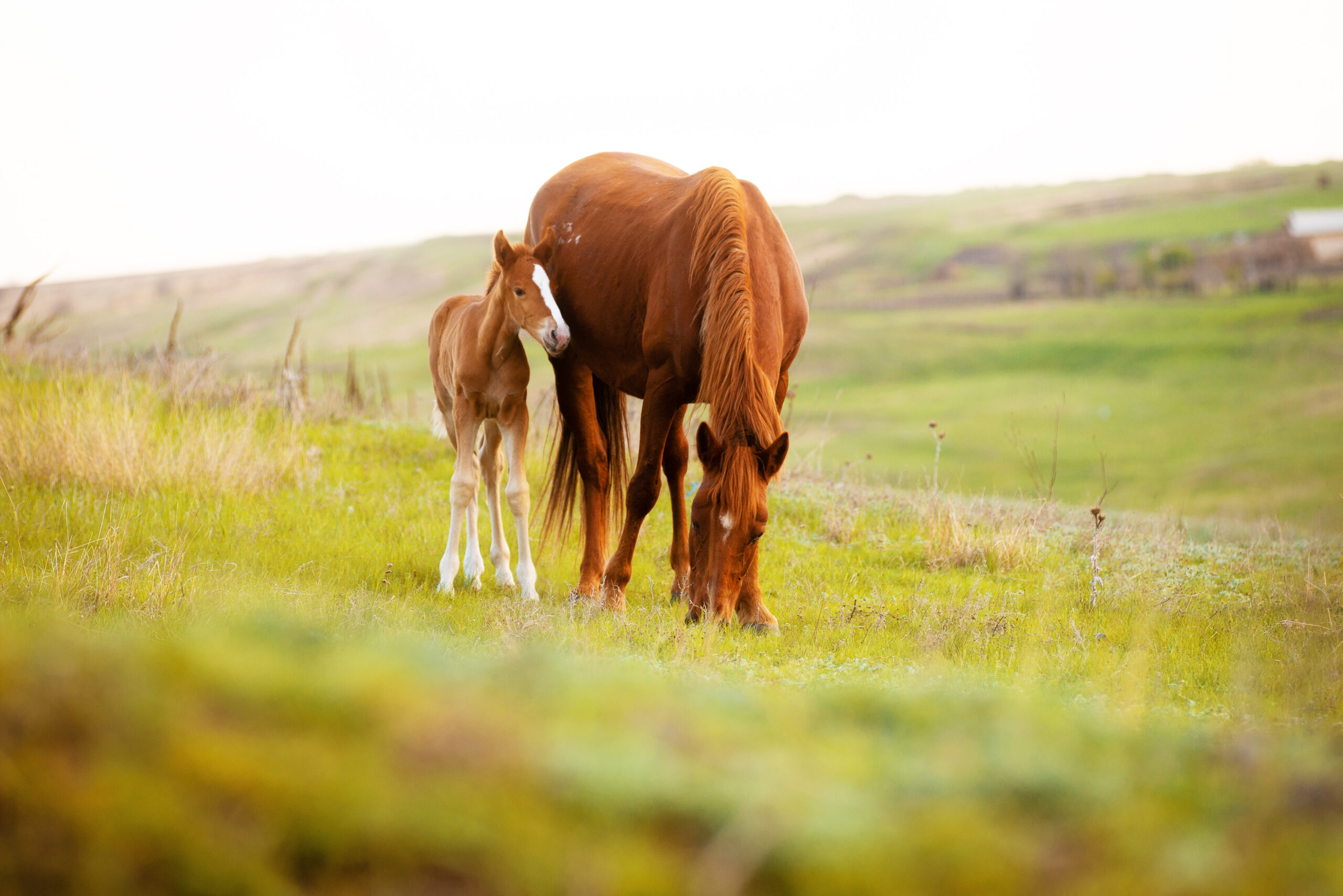
By the time the June issue reaches readers, the regular breeding season is winding down for many horse owners. Some mares are already confirmed in foal, some breeding decisions have been pushed aside for another year and some owners are already thinking ahead to next season. For those with an older mare they hope to breed, that early planning can make a real difference.
Older mares can still produce foals, but they may need more help than younger mares. Age affects the reproductive system, and those changes can make it harder for a mare to become pregnant, stay pregnant or carry a healthy foal to term. That does not mean every older mare is a poor candidate for breeding. It does mean owners should go into the process with realistic expectations and a good veterinarian involved from the beginning.
A mare’s reproductive age does not always match her number of birthdays. Some mares in their mid-to-late teens settle easily and carry foals without much trouble. Others begin having problems earlier. Past reproductive history matters. A mare that has had regular foals may be different from an older maiden mare that spent most of her life showing, racing, working or sitting open. Health, body condition, uterine health and conformation all play a role.
One of the main challenges with older mares is egg quality. As mares age, their eggs age, too. Older eggs are more likely to have abnormalities that prevent fertilization, stop early embryo development or lead to early pregnancy loss. Even when breeding is timed well and the stallion has good fertility, the mare may not settle because the egg itself is no longer as viable as it once was.
The uterus also changes with age. The lining of the uterus, called the endometrium, can become less healthy and less able to support pregnancy. Over time, some mares develop fibrosis or scarring within the uterus. This can interfere with the placenta’s ability to develop and support a growing fetus. A mare may get pregnant early, but then lose the pregnancy because the uterus cannot maintain it.
Older maiden mares can face an additional problem. When a mare cycles, it is normal for some fluid to be present in the uterus. A healthy reproductive tract clears that fluid through uterine contractions and cervical relaxation. In some older mares, especially those that have never had a foal, the cervix may not relax well enough. Fluid can build up and remain in the uterus after breeding. That fluid can damage sperm, interfere with an embryo and increase the risk of infection or inflammation.
Post-breeding inflammation is another concern. All mares experience some uterine inflammation after breeding, but most clear it within a reasonable amount of time. Older mares may not clear it well. This can leave the uterus in a poor environment for pregnancy. A veterinarian may recommend uterine lavage, medication or other treatment after breeding to help the mare clear fluid and inflammation.
External conformation can also affect fertility. As some mares age, especially taller, thinner mares or mares that have had multiple foals, the reproductive tract may tilt in a way that allows manure or air to contaminate the vulva and vagina. Poor vulvar conformation can increase the risk of uterine infection. In some cases, a veterinarian may recommend a Caslick’s procedure to help protect the reproductive tract.
Because so many factors can be involved, an older mare should have a breeding soundness exam before the next season begins. This may include a physical exam, reproductive ultrasound, uterine culture, cytology and possibly a uterine biopsy. These tools help determine whether the mare has infection, inflammation, fluid retention, poor uterine health or other issues that need to be addressed before breeding.
Timing is also important. With an older mare, it is usually better to start early in the season rather than waiting until late. More cycles give the veterinarian more chances to manage the mare properly. Early planning also allows time to treat infection, improve body condition, schedule semen shipments, evaluate the stallion’s fertility and consider whether cooled semen, frozen semen, live cover or another option makes the most sense.
Owners should also look closely at the mare’s overall health. A mare that is too thin, too heavy, metabolically unstable, lame or dealing with chronic illness may struggle to conceive or carry a foal. Good nutrition, dental care, hoof care and vaccination planning all matter. Breeding may be a reproductive decision, but pregnancy affects the whole horse.
For some mares, advanced reproductive options may be worth discussing. Embryo transfer allows a valuable mare to produce a foal without carrying it herself. This may be helpful for older mares that can produce an embryo but should not carry a pregnancy, or for mares that are still competing. Other assisted reproductive techniques may also be available through specialized equine reproduction centers.
The hard truth is that breeding an older mare can take more time, money and patience. Not every mare will get in foal, even with good care. Some mares should not be bred if the risks are too high. Still, many older mares can produce healthy foals when problems are identified early and managed correctly.
For owners hoping to raise one more foal from a favorite mare, the best plan is to start the conversation before breeding season arrives. Talk with a veterinarian, evaluate the mare honestly and make decisions based on her health, history and reproductive exam. Hope is part of breeding horses, but planning gives that hope a much better chance.
By Summer McMillen
Everyone that knows anything about horses knows that there are bad ones, good ones, and great ones.
The bad ones are good for nothing. You can’t catch them, you can’t saddle them, and you can’t get on them without feeling like you need a helmet, some kind of padded vest and, an instruction manual. Once you do mount up the whole ride is a battle and heaven forbid, you actually have a job to do because they are little to no help in holding the herd. We all find ourselves owning a bad one or two throughout our lives. Best case scenario is they find a more tolerable home to go to through via a horse sale or the classifieds. Worst case scenario all you can do is say “Vaya con Dios,” put a sign on them that reads “Do Not Attempt,” and turn them out to pasture. Hoping they are decent enough to stay within the borders and make a beautiful yard ornament.
Good horses are usually much more tolerable. They’re pretty easy to catch, saddle, and hop up on. Sometimes they might have a bad habit or two like setting back when they’re tied to a fence or, getting cold backed on early mornings that you tolerate because they are so skilled in a specific field. A good horse is usually only good for one thing. They have a niche talent m, if you will. They can be a good heel horse. A good head horse. The horse you want to gather pastures on because you know he won’t knicker or rare up when you get dropped off in the jig line. A good kid horse. Your rodeo horse. The horse you put your wife on when she’s being a little wimpy that day. Good horses usually get sold because they are proficient in their given field and they find good homes making both parties happy. We will all own many good horses in our lives and be happy to do so.
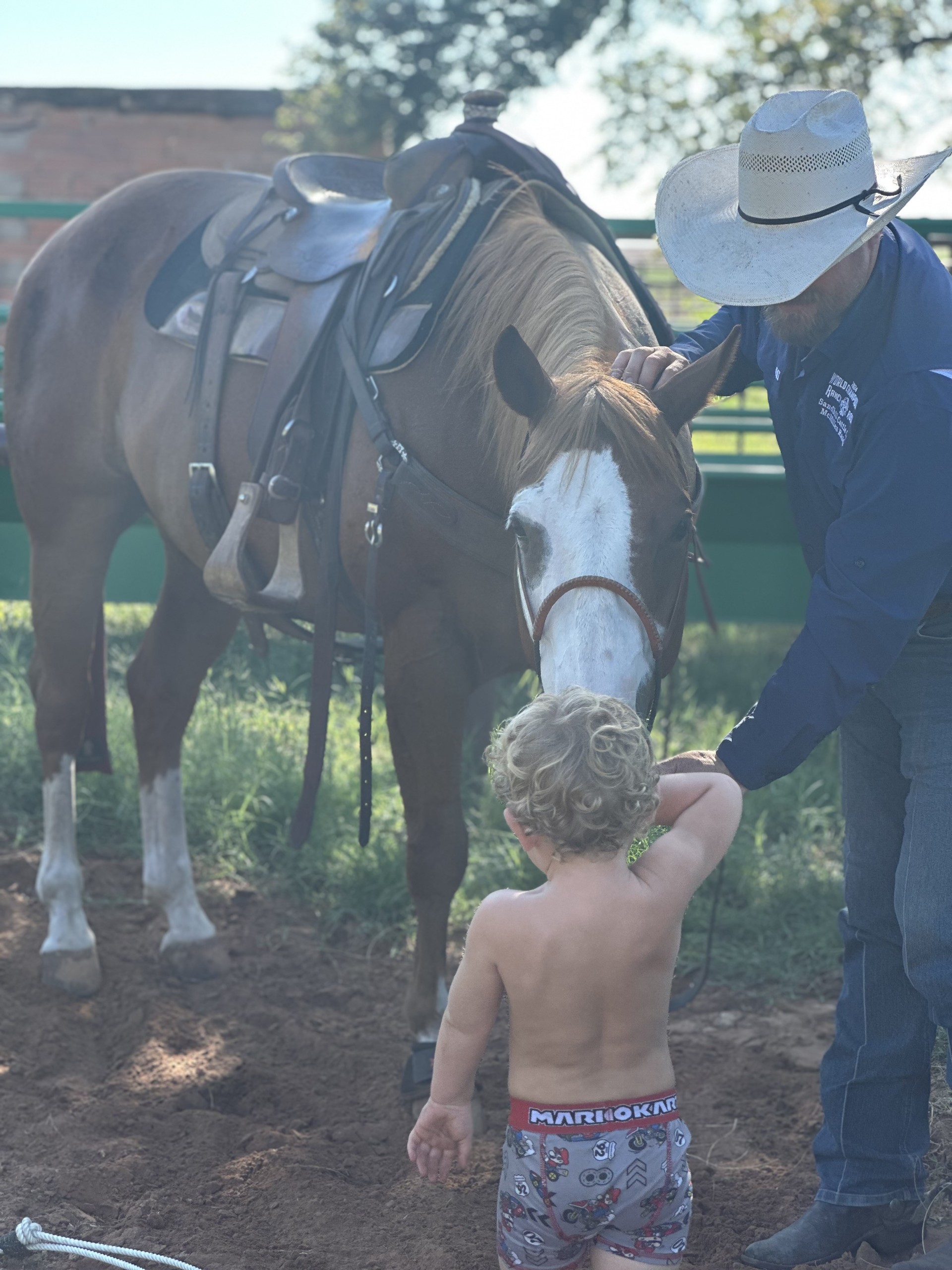
Great horses are a rare and treasured possession. They are simultaneously easy and hard to own. Easy because you can do anything on them. Hard because everyone is always trying to buy them from you. A great horse stands still while your kid pulls their head down all the way to the ground so they can halter them. A great horse is never cold backed and always ready to cinch tight and take off. A great horse can be ridden in the pasture and the rodeo arena on the same day. A great horse doesn’t need practice. A great horse is always willing to do anything you ask of them at any given moment. Great horses find their homes as horse colts and usually live out the rest of their days at the same home because great horses are irreplaceable.
People and horses are not all that different. There are bad, good, and great ones. The more time I spend around horses the more I am convinced of the kind of person I want to be. “Bad” will absolutely not do. “Good“ is much too common and just doesn’t quite cut it more often than not. “Great” is what I aspire to be.
Great can be defined in so many ways when we let human standards get involved but, I want to be great as defined in the qualities of a great horses.
I want to be kind and patient while my children are learning. I want to be ready to help anyone who asks me. I want to go the extra mile. I want to make my home a beautiful place to come to after a full day’s work outside. I want to not be thrown off by life’s twists and turns but, firm in my faith.
So, basically what I’m saying is I want to be a great horse. And honestly there are worse things we could all aspire to be.
Here’s to great horses. May we know them, love them, and if we’re lucky be great just like them.


-
 Country Lifestyle2 years ago
July 2017 Profile: J.W. Hart
-
 Attractions9 years ago
48 Hours in Atoka Remembered
-
 Farm & Ranch2 years ago
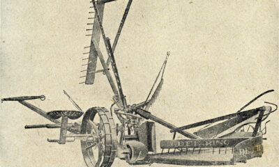
From Plow to Plentiful: The Most Important Inventions in Agricultural History
-
 Equine9 years ago
Umbilical Hernia
-
 Outdoors8 years ago
Grazing Oklahoma: Honey Locust
-
 Country Lifestyle5 years ago
The Two Sides of Colten Jesse
-
 Farm & Ranch8 years ago
Hackberry (Celtis spp.)
-
 Equine6 years ago
On the Road with Emily Miller-Beisel