Equine
Equine Flexural Limb Deformities
By Dr. Garrett Metcalf
Flexural limb issues can occur in different age groups of horses, starting with newborns up to two- to three-year-olds. These issues occur somewhat predictably in age groups and can be addressed rather quickly when needed. There are various treatments and methods that can be used to address flexural issues. This article will discuss the most common flexural abnormalities and treatment methods.
Foal Flexural Issues
Foal flexural issues are often considered congenital flexural limb abnormalities because they are born with them. We don’t fully understand why this occurs but there is some evidence in the human literature that lack of fetal activity in the womb causes club feet in babies. In foals, it is thought that uterine positioning is to blame for part of the contracted tendons. Other causes can be exposure of the mare to toxic plants or substances that may be toxic to the fetus.
The most common area that a foal will have contracture of limb is at the carpus or knee. These foals will not be able to fully extend the knee and often will affect both at the same time. These foals can have difficulty standing to nurse or will get fatigued quickly and will not be able to stand for longer periods of time. There can also be damage to the extensor tendons or even rupture of extensor tendons caused by the high strain placed on them when the foal tries to stay standing. The rupturing of these tendons is not overly concerning but the lack of extensor function can make the flexural limb deformity worsen.
Other common locations of flexural limb deformities can be at the fetlock or coffin joint level. These deformities are not usually as detrimental to allowing the foal to stand and nurse properly compared to carpal flexural deformities. These deformities can be addressed similar to carpal deformities with some exceptions.
Treatment of Flexural Deformities
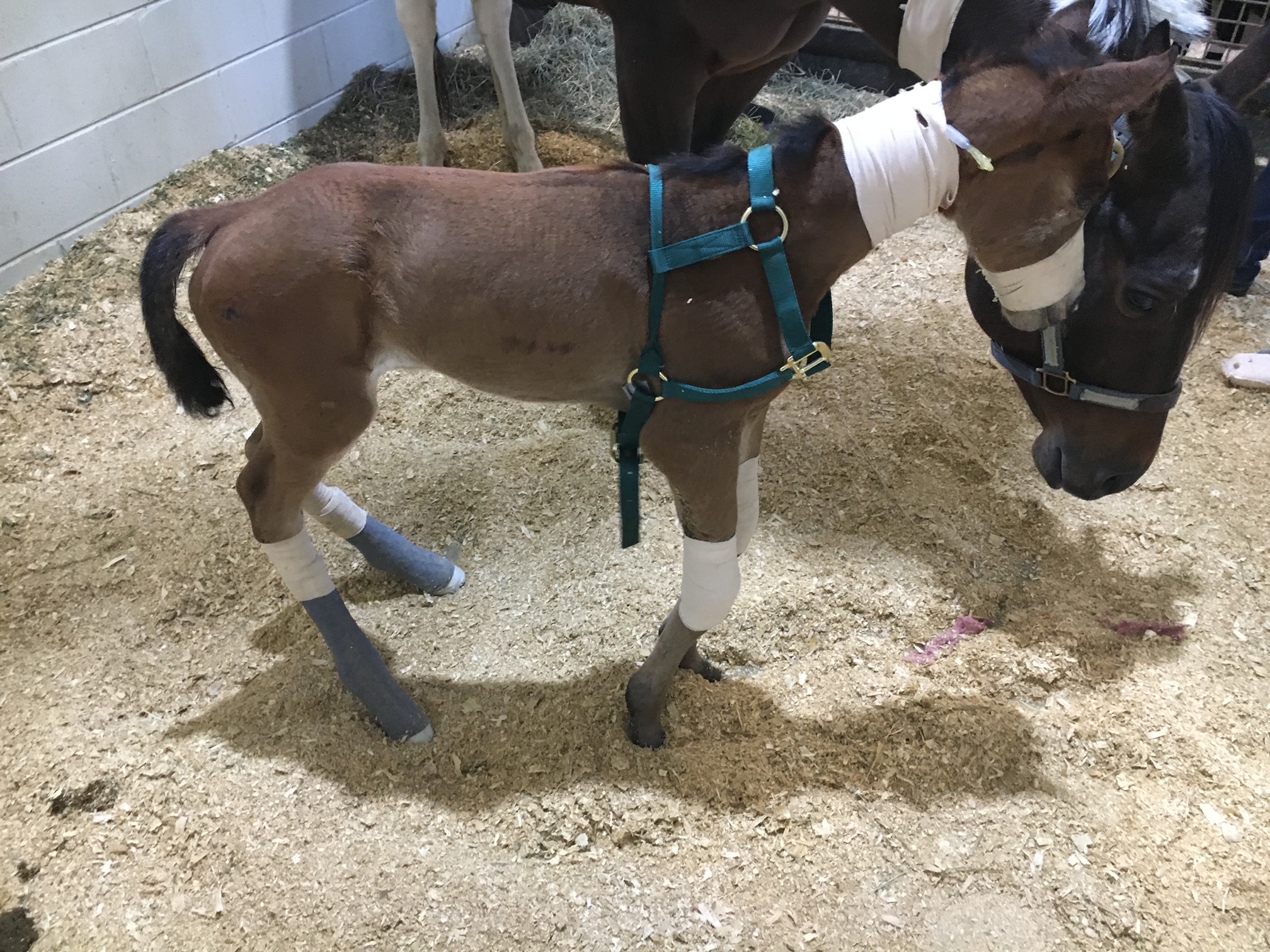
Splints or casts can be used to stretch and support the effected limbs of foals. Splints are often preferred by most veterinarians because they can be repositioned or reset as needed. Splints are easier to place on the limbs of foals but they do need resetting every 24 to 48 hours. Casting of the limbs is more rigid but is not adjustable once placed. Casting is often needed in more severe cases and requires changing frequently. Whenever placing these devices, care must be taken to prevent splint or cast sores because foal skin is rather delicate.
Surgical intervention is needed in some cases of carpal flexural deformities. A study out of Australia found that cutting of two muscle/tendon groups on the back of the carpus greatly improved the ability to extend the carpus with splinting methods. Cutting of these tendons do not have consequence to future athletic function. The two muscles are called flexor carpi ulnaris and ulnaris lateralis.
An antibiotic called Oxytetracycline is helpful to treat flexural limb deformities because of its side effect of causing tendon laxity. The laxity is created by chelating calcium within the tendons and allows the relaxation of tendons. This method does have some risk because of the high dose required and renal injury that it can cause when not administered with IV fluids.
Toe extension shoes are used when it comes to dealing with lower limb flexural limb deformities. These shoes are often applied with adhesives and after the splinting or casting is no longer needed. The toe extension shoe allow foal to continue to stretch those tendons every time they take a step and prevent from becoming contracted again.
Older horses (six months or older) with contracted tendons often get acquired limb deformities and the horses need surgical intervention to correct these deformities. These surgeries cut or release check ligaments that allows the musculotendinous unit of the deep digital or superficial digital flexor tendon to elongate. The deep digital flexor tendon is responsible for causing club feet or a flexural limb deformity at the coffin joint. The superficial digital flexor tendon is responsible flexor tendon that causes a flexural limb deformity at the fetlock joint. The check ligaments attach the tendon to bone and do not allow the tendon to elongate past a certain point. By eliminating these ligaments the flexural limb deformity can be corrected by allowing the muscle to stretch since the tendon is much more rigid.
Flexural limb deformities can be caused by excessive laxity or weakness of the tendons. These deformities are often seen in premature foals or foals that are born at a much smaller birth weight. The excessive laxity will cause the toes of there feet to flip up in the air and the fetlocks to be touching the ground. The areas where the skin is contacting the ground will cause sores and abrasions. If these areas are note protected the wounds can get into deep structures causing serious infection and injury the flexor tendons.
Treatment for tendon laxity is to add heel extension shoes to keep the toes flat to the ground. The extension behind the foot forces the toe down under the foals own weight. As the foal becomes stronger from normal activity the muscle attached to the tendons can support the foal and the limb laxity will correct itself. Abrasions still can occur even with heel extension shoes are in place so bandages need to be applied to protect these areas.
Flexural limb issues are a common issue that horses and owners will face. It is best to have your horse evaluated by a veterinarian whenever these problems are suspected. Foal flexural limb deformities can be life threatening because of the limitation of standing on time to nurse colostrum. Without colostrum within the first hours of life the foal is a much higher risk of sepsis and death.
Read more in the August 2023 issue of Oklahoma Farm & Ranch.
By Summer McMillen
Everyone that knows anything about horses knows that there are bad ones, good ones, and great ones.
The bad ones are good for nothing. You can’t catch them, you can’t saddle them, and you can’t get on them without feeling like you need a helmet, some kind of padded vest and, an instruction manual. Once you do mount up the whole ride is a battle and heaven forbid, you actually have a job to do because they are little to no help in holding the herd. We all find ourselves owning a bad one or two throughout our lives. Best case scenario is they find a more tolerable home to go to through via a horse sale or the classifieds. Worst case scenario all you can do is say “Vaya con Dios,” put a sign on them that reads “Do Not Attempt,” and turn them out to pasture. Hoping they are decent enough to stay within the borders and make a beautiful yard ornament.
Good horses are usually much more tolerable. They’re pretty easy to catch, saddle, and hop up on. Sometimes they might have a bad habit or two like setting back when they’re tied to a fence or, getting cold backed on early mornings that you tolerate because they are so skilled in a specific field. A good horse is usually only good for one thing. They have a niche talent m, if you will. They can be a good heel horse. A good head horse. The horse you want to gather pastures on because you know he won’t knicker or rare up when you get dropped off in the jig line. A good kid horse. Your rodeo horse. The horse you put your wife on when she’s being a little wimpy that day. Good horses usually get sold because they are proficient in their given field and they find good homes making both parties happy. We will all own many good horses in our lives and be happy to do so.
Great horses are a rare and treasured possession. They are simultaneously easy and hard to own. Easy because you can do anything on them. Hard because everyone is always trying to buy them from you. A great horse stands still while your kid pulls their head down all the way to the ground so they can halter them. A great horse is never cold backed and always ready to cinch tight and take off. A great horse can be ridden in the pasture and the rodeo arena on the same day. A great horse doesn’t need practice. A great horse is always willing to do anything you ask of them at any given moment. Great horses find their homes as horse colts and usually live out the rest of their days at the same home because great horses are irreplaceable.
People and horses are not all that different. There are bad, good, and great ones. The more time I spend around horses the more I am convinced of the kind of person I want to be. “Bad” will absolutely not do. “Good“ is much too common and just doesn’t quite cut it more often than not. “Great” is what I aspire to be.
Great can be defined in so many ways when we let human standards get involved but, I want to be great as defined in the qualities of a great horses.
I want to be kind and patient while my children are learning. I want to be ready to help anyone who asks me. I want to go the extra mile. I want to make my home a beautiful place to come to after a full day’s work outside. I want to not be thrown off by life’s twists and turns but, firm in my faith.
So, basically what I’m saying is I want to be a great horse. And honestly there are worse things we could all aspire to be.
Here’s to great horses. May we know them, love them, and if we’re lucky be great just like them.
By Dr. Garrett Metcalf
The suspensory ligament is a vital component of the limb of a horse to produce normal locomotion and support. The suspensory ligament is a common area of concern in performance horses of various disciplines and can be single handedly the cause of lameness or performance issues. This article is going to look at a specific degenerative disease of the suspensory ligament and what horses are at risk for this disease.
DSLD or degenerative suspensory ligament desmitis was first discovered in the early 1980’s in Peruvian Paso horses. The name has been changed because the suspensory ligament is not the only organ affected from the disease but the suspensory is ultimately the biggest issue. The newer name, ESPA or equine systemic proteoglycan accumulation, is more correct because other ligaments and tissues are affected by this disease. In this article we will only focus on the suspensory ligament. The most commonly affected breeds are Peruvian Paso, Paso Fino, Morgan, Saddlebred, Warmblood, Paints, American Quarter Horse, and Thoroughbred breeds. The age of onset of the disease is variable among breeds but it is more common to be seen in middle age to older horses. However it has been documented in horses as young as one year of age. The disease generally will have a slow insidious onset that can go undiagnosed for months or years depending on the horses work and discipline.
A horse that begins to show early signs of DSLD may have a vague lameness issue that is difficult to isolate and they most likely will resolve with a period of rest. As the horse returns to moderate level of work the lameness will return. This scenario may go on for several months or more before the discovery of the DSLD is made. The first indication of DSLD is often pain isolated in the suspensory branches or fetlock region when a flexion test is performed. Horses with DSLD will also have a “dropped” fetlock appearance because the suspensory is the main supporting structure of the fetlock joint. DSLD can affect the hind limbs, forelimbs or all limbs at the same time. A unique sign of DSLD is that not just one limb is affected but rather bilaterally affecting the limbs, meaning it will either affect either both forelimbs or hind limbs at the same time. It is my experience that the hind limbs are more commonly affected compared to the forelimbs. Horses will often have enlargement of the fetlock region and increased joint fluid or wind puffs. Horses will often have a straight hock or post legged hind limb appearance. Horses will often shift weight frequently in an effort to get relief from the discomfort and this can be confused with other lameness issues or foot related pain.
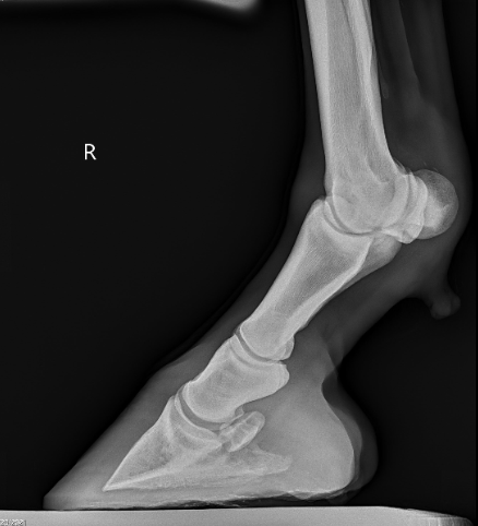
Diagnosis of DSLD is often made by clinical signs, breed and ultrasound findings. Ultrasound imaging of the suspensory ligaments will often show diffuse enlargement of the suspensory body and branches. The suspensory ligament will have a poor heterogeneous fiber pattern with periligamentious soft issue thickening from scar tissue deposition and edema or fluid within the tissue. Radiographs of the lower limb may reveal abnormal bone changes in the sesamoid bones behind the fetlock joints and even osteoarthritis of the pastern and or fetlock joints. A definitive diagnosis can be made from a biopsy of a ligament in the neck called the nuchal ligament, but is not often performed because of the invasiveness of the biopsy.
Treatment is very limited and it is mostly geared towards protection of further damage by prolonged rest. Pain management is also important to attempt to keep the horse as comfortable as possible. Different shoeing techniques can be used with marginal success. In early cases of DSLD, a suspensory shoe that helps engage more work from the deep digital flexor tendon can help elevate the fetlock and offer more protection to the suspensory ligament. The devastating thing about this disease is that there is no cure and there are hardly any good options to slow the progression of the disease. DSLD carries a poor prognosis when the diagnosis is made in any breed of horse or any discipline. Although some cases can be managed better than others, it often progresses to the point of debilitating pain and discomfort to the point of humane euthanasia especially in the Peruvian Paso breed.
Read more in the February 2023 issue of Oklahoma Farm & Ranch.
By Dr. Devan England DVM
Does your horse have gastric ulcers? Gastric or stomach ulcers are frequently blamed for a variety of things including poor performance, acting ‘cinchy’, weight loss, not eating, poor coat condition, diarrhea and colic. However, gastric ulcers are not always the culprit and the only way to know for sure if your horse has gastric ulcers is to look at the stomach on camera, using an endoscope. Poor appetite and poor body condition are the mostly widely observed clinical signs with gastric ulcers, however, these are non-specific. If you think your horse might have gastric ulcers, the best place to start is to talk to your veterinarian and consider scheduling a gastroscopy. Gastroscopy requires the horse be held off feed for at least 16-18 hours and held off water for at least 6-8 hours. Fasting off feed and water is necessary to allow the veterinarian to see the whole stomach. If restricting feed or water is difficult in your management situation, many veterinarians will allow you to hospitalize your horse the night before gastroscopy for proper fasting.
Gastric ulcers are split into two types, classified by the location of the ulcer in the stomach. Squamous ulcers are ulcers that occur in the squamous or skin like portion of the stomach. This is the top part of the horse’s stomach, is closest to the esophagus, and has squamous tissue to protect this portion of the stomach from stomach acids. The other ulcer type are glandular ulcers. Glandular ulcers occur in the bottom portion of the stomach, which is closest to the small intestine. This portion of the stomach has glandular mucosa with cells responsible for producing stomach acids for digestion as well as cells that produce mucus and buffers to protect the lining from stomach acid. Gastroscopy is important not only for diagnosing whether ulcers are present but also determining the severity and the type of ulcer, because these two ulcer types require different treatments.
Squamous gastric ulcers are common in racehorses both in and out of training, with higher prevalence in racehorses under training. Prevalence in Thoroughbred racehorses in training has been reported to be up to 100% (Sykes 2015). Squamous ulcers are also prevalent in Western pleasure horses, Thoroughbred stallions on breeding farms, and Italian donkeys (Sykes 2015). Glandular gastric ulcer prevalence has not been as well described as squamous ulcers. Glandular ulcers are reported to be most common in Thoroughbred and Standardbred racehorses, Canadian showjumpers and polo ponies, and American Quarter Horses (Sykes 2015).
Risk factors for ulcers vary by ulcer type. Anti-inflammatories (Bute, Banamine) can increase the risk of glandular ulcers in some horses by affecting normal defense mechanisms but are not a high risk in most horses. Horses that display stereotypic behaviors, such as cribbing, have an increased risk of squamous ulcers. Grain fed before hay in non-exercising horses, feeding larger amounts of grain, and increased time between meals increases the risk of squamous ulcers. Increased time with high intensity exercise and housing in single pens is associated with increased risk of glandular ulcers. A straw only diet, lack of water access and lack of direct contact with other horses increases the general risk of gastric ulcers.
If your horse is diagnosed with ulcers, the mainstay of treatment is a buffered formulation of omeprazole (Gastrogard, Ulcergard). Over the counter Omeprazole and compounded Omeprazole are not effective because without buffering, the acidic stomach quickly breaks down the drug before absorption. Most horses with squamous ulcers will have healing of these ulcers after a 4-week course of Gastrogard or Ulcergard at treatment dose (whole tube for the average horse). Some horses may be healed by 3 weeks of treatment, but all horses should undergo a recheck gastroscopy before stopping treatment. Horses diagnosed with glandular ulcers need combination therapy with Gastrogard/Ulcergard and Sucralfate for 4 weeks. About 2/3 of horses with glandular ulcers will heal in this time, but some horses may require longer treatment times so a recheck is always recommended before discontinuing treatment.
Horses at higher risk of gastric ulcers may benefit from preventative (low) doses of Ulcergard (1/4 tube in average sized horse) given for a few days before and during high stress situations like long distance travel and competitions. Sea buckthorn berry supplement may be protective against formation of glandular ulcers. Dietary management to decrease the risk of ulcers includes providing more frequent small hay meals if pasture access is not available, limiting high sugar grains as much as possible and adding vegetable oil to the feed.
Sykes BW, Hewetson M, Hepburn RJ, Luthersson N, Tamzali Y. European college of equine internal medicine consensus statement – equine gastric ulcer syndrome in adult horses. J Vet Internal Med 2015; 29:1288-1299.


-
 Country Lifestyle2 years ago
July 2017 Profile: J.W. Hart
-
 Attractions9 years ago
48 Hours in Atoka Remembered
-
 Equine9 years ago
Umbilical Hernia
-
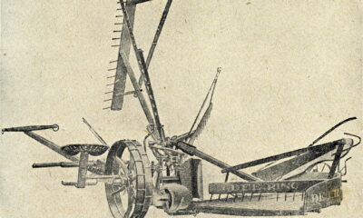
 Farm & Ranch1 year ago
From Plow to Plentiful: The Most Important Inventions in Agricultural History
-
 Outdoors8 years ago
Grazing Oklahoma: Honey Locust
-
 Country Lifestyle5 years ago
The Two Sides of Colten Jesse
-
 Farm & Ranch8 years ago
Hackberry (Celtis spp.)
-
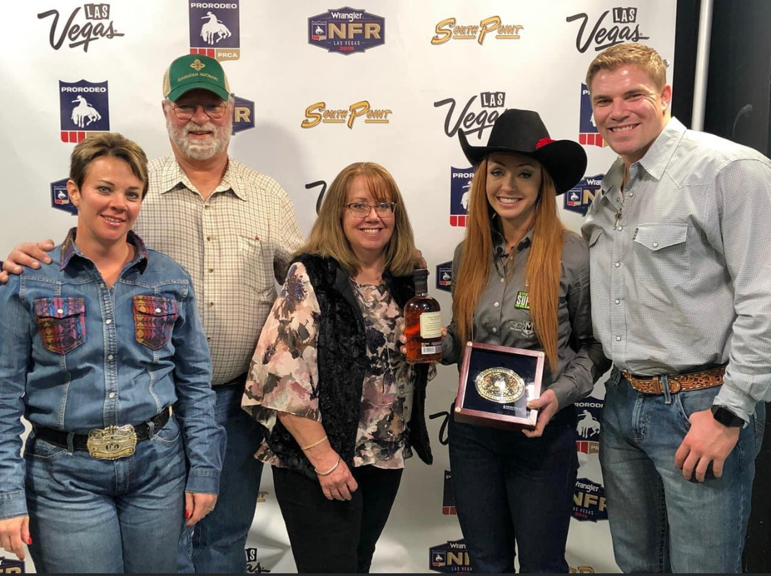 Equine6 years ago
On the Road with Emily Miller-Beisel