Farm & Ranch
African Swine Fever (Update)
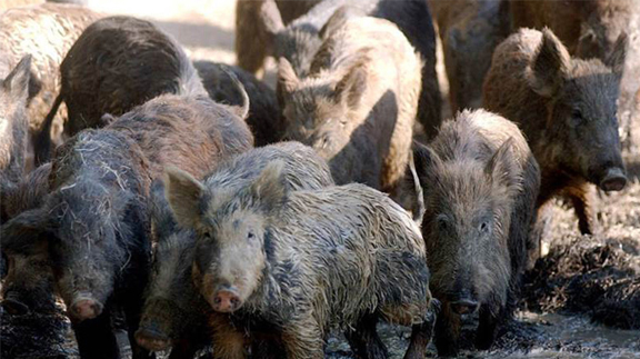
On July 28, 2021, the United States Department of Agriculture (USDA) confirmed African Swine Fever (ASF) in the Dominican Republic. The USDA is assisting the Dominican Republic with their efforts to contain and control this virus as well as offering help to Haiti which borders the Dominican Republic. The Dominican Republic is slightly over 200 miles from Puerto Rico. With Puerto Rico being a territory of the United States (US), any ASF found there would result in the World Organization for Animal Health (OIE) restricting the exportation of pork from the US. For this reason, the USDA is establishing a Foreign Animal Disease protection zone in Puerto Rico and the US Virgin Islands and is increasing efforts to keep ASF out of the continental US.
Loss of movement of pork from the US would have significant economic consequences in the US and Oklahoma. According to Kylee Deniz executive director of the Oklahoma Pork Council, Oklahoma pig farmers generate $5.7 billion in annual revenue. Loss of exports would not only affect pig producers but beef and poultry producers as well. All swine producers whether commercial, show pig producers, or kids with one 4H show pig must protect the Oklahoma pig industry. All pig producers should have a biosecurity protocol in place as well as be familiar with the symptoms of ASF.
The African Swine Fever virus is in the Asfarviridae family. The virus infects domestic and feral swine. The virus is found in all pig secretions especially oronasal fluids of infected swine. The virulence of the virus varies considerably between strains with some strains resulting in large number of deaths and some with little sickness at all. The virus is resistant. Most common disinfectants will not destroy the virus. It will survive for long periods of time in blood, soil, and uncooked pork products.
The virus is easily transmitted by direct contact, indirect contact, and insect vectors. It may spread from pig to pig by inhaling the virus. Other ways that pigs may be directly infected are by ingesting the virus in uncooked pork products or by cannibalism. Fomites such as vehicles, footwear, clothing, equipment, and feed may serve as ways to introduce the virus to a farm. Feed and feed ingredients are especially worrisome. In simulated Trans-Atlantic and Trans-Pacific shipping models, ASF virus survived in feed ingredients (Dee et al.,2018). These experiments simulated shipping feed ingredients from Asia and Europe to the US. Lastly, the Ornithodoros ticks which are soft ticks are known to harbor the virus for long periods of time. These insects transmit the virus to pigs.
Once a pig is infected with the virus, clinical signs will usually appear in 5 to 21 days. Sudden death may be the only clinical sign seen. Milder cases are often confused with other pig diseases. Clinical signs often observed are high fever, loss of appetite, weakness, and recumbency. Skin lesions sometimes seen are red blotchy areas or blackened areas. Infected pigs will have trouble breathing. Ocular and nasal discharges are seen in some pigs. Digestive signs include diarrhea, vomiting, and constipation. Pigs tend to have bleeding episodes such as nose bleeds or bloody diarrhea. Pregnant animals tend to abort. Abortion may be the first sign of the disease. Even though some strains of the virus cause minor clinical signs, most strains result in large numbers of sick pigs with several of them dying.
Treatment is not an option with ASF. Any swine operation that is found to have an ASF outbreak will be forced to depopulate and go through a rigorous cleaning and disinfecting of the farm. Vaccine research is ongoing, but no USDA licensed vaccine is available in the US at this time which means producers must rely on a good biosecurity plan to protect their premises.
The USDA is working hard to keep ASF out of the US. However, many veterinarians would agree that if ASF were to be found in the US and the virus infected the feral pig populations, it would be very difficult to eradicate the virus from the US. For this reason, all swine producers need to focus on their disease prevention strategies to protect their animals. If producers would like more information about ASF, they should review the Foreign Animal Disease Prevention & Preparedness at www.pork.org/FAD or contact their local veterinarian or local Oklahoma State University County Agriculture Extension Educator.
References
Dee SA, Bauermann FV, Niederwerder MC, Singrey A, Clement T, de Lima M, Long C, Patterson G, Sheahan MA, Stoian AMM, Petrovan V, Jones CK, De Jong J, Ji J, Spronk GD1, Minion L, Christopher-Hennings J, Zimmerman JJ, Rowland RRR, Nelson E, Sundberg P, Diel DG . Survival of viral pathogens in animal feed ingredients under transboundary shipping models. PLoS One. 2018 Mar 20;13(3):e0194509.
Farm & Ranch
Fescue Foot
Barry Whitworth, DVM | Area Food/Animal Quality and Health Specialist for Eastern Oklahoma
*Article originally printed in the October 2022 issue of Oklahoma Farm & Ranch.
Since most of Oklahoma experienced drought conditions and with fall fast approaching, producers with fescue pastures should closely observe their livestock for any signs of fescue toxicity. According to Mike Trammel, Pottawatomie County Ag Educator and Muti-County Agronomist, fescue toxins (ergot alkaloids) tend to increase in Kentucky-31 tall fescue pastures in the fall. Some reports indicate more problems with fescue toxins following a summer drought and limited fall rains. All of this may put Oklahoma cattle at a greater risk of fescue toxicity.
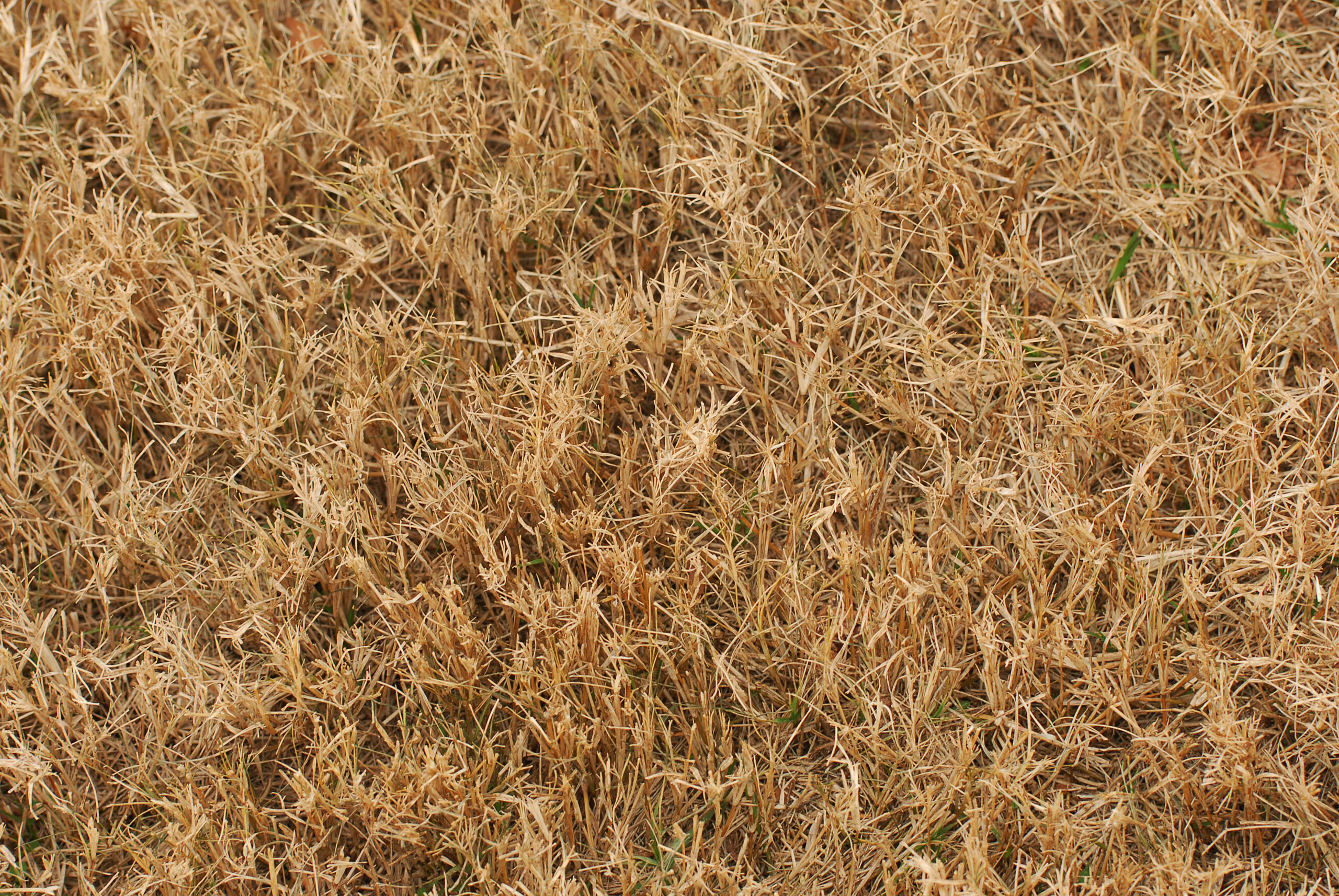
One issue that cattle experience with fescue toxins is fescue foot. Fescue foot is thought to be caused by ergot alkaloids such as ergovaline. These alkaloids are produced by endophyte fungus (Epichloë coenophiala) which is in tall fescue. Ergovaline has been proven to be a vasoconstrictor which might be responsible for fescue foot and heat intolerance also known as summer slump in cattle. Other issues that may be seen with the ergot fescue toxins are reduced milk production and reproductive issues.
Clinical signs of fescue foot appear within a few days of cattle being turned on to tall fescue pastures or it may take weeks if toxins in the pasture are low. Producers will initially observe cattle with arched back, rough hair coats, and sore feet. These symptoms are more noticeable early in the morning and with cold weather. This is followed by reddening and swelling in the area between the dewclaws and hooves. The lameness usually becomes more severe with time. If no action is taken, gangrene will result in loss of tissues distal to the coronary band and declaws. If the weather remains mild, other signs such as increase respiration rate, increase heart rate, and higher body temperature are more common.
Other causes of lameness in cattle must be differentiated from fescue foot. One simple method that will help differentiate fescue foot from footrot is to check the temperature of the foot. If the foot is cold, this is an indication that the problem is more likely fescue foot.
Since there is not a specific treatment for fescue foot, the condition must be managed. Cattle need to be observed daily for any signs of lameness or stiffness during the first few weeks on fescue pastures. This should be done early in the morning before cattle walk off the stiffness. Producers should pay close attention during cold weather, especially when rain, snow, or ice are present. Any animal showing clinical signs of fescue foot should be removed from the pasture and placed in a clean environment. The animal should be fed a ration with no fescue toxins.
The best but most costly solution to reduce fescue toxicity is to renovate old pastures with new endophyte friendly varieties. If this option is not possible, producers might try interseeding fescue pastures with clovers or other grasses. This should dilute fescue toxins. Nitrogen fertilization may increase ergot alkaloids, so producers should avoid fertilizing fescue pastures with high amounts of nitrogen. Researchers have demonstrated that feeding a supplement while grazing fescue pastures reduces clinical symptoms. Some studies indicate a difference in susceptibility to fescue toxicity in some cattle. Selecting cattle based on genetic tolerance of fescue toxins is an option. (For more information go to www.agbotanica.com/t-snip.aspx)
With large areas in Oklahoma covered with Kentucky-31 fescue pastures, fescue foot as well as other fescue toxicities are not going away any time soon. Livestock producers will need to watch their livestock closely for any signs of fescue toxicity and manage their pastures to keep toxins as low as possible. If producers would like more information on fescue foot, they should consult their veterinarian and/or visit their local Oklahoma State University Cooperative County Extension Agriculture Educator.
Farm & Ranch
Animal Disease Traceability
Barry Whitworth, DVM, MPH | Senior Extension Specialist | Department of Animal & Food Sciences | Freguson College of Agriculture
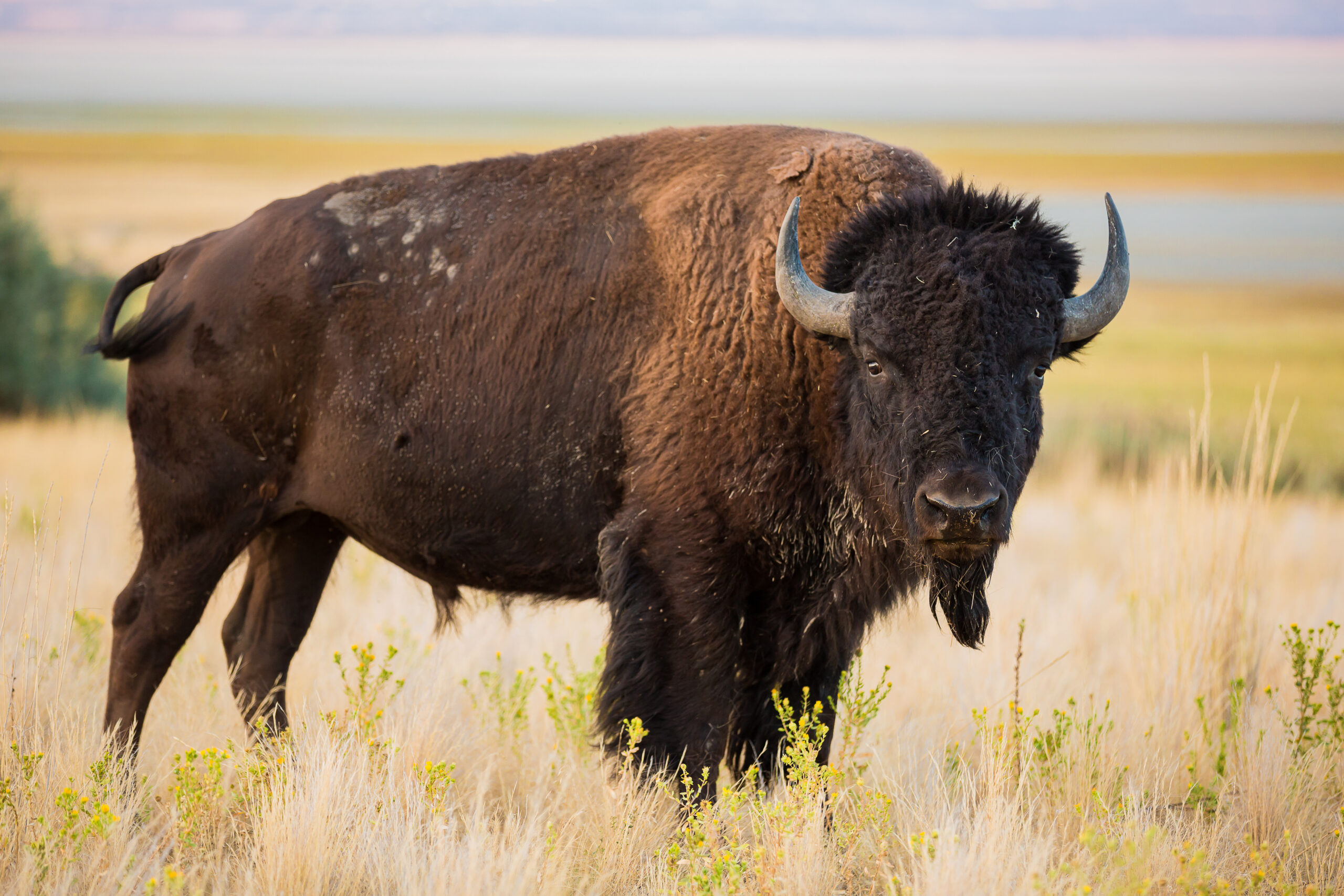
On July 6, 2020, the United States Department of Agriculture (USDA) Animal and Plant Health Inspection Service (APHIS) posted in the Federal Register a proposal that radio frequency identification tags be used as official identification for cattle and bison. Following a period for public comment, the USDA APHIS released a statement on April 24, 2024, with the amended animal disease traceability (ADT) regulation for cattle and bison. The full press release may be found at https://www.aphis.usda.gov/news/agency-announcements/aphis-bolsters-animal-disease-traceability-united-states. Under the new rule, cattle and bison will need to be identified with tags that are both visual and electronic.
The USDA defines ADT as knowing where diseased and at-risk animals are, where they have been, and when the animal disease event took place. A system that allows for efficient traceability of livestock in the United States (US) is essential for animal health and reducing the economic effect of a foreign animal disease outbreak and other diseases on livestock producers as well as others whose well-being depends on livestock production.
In the past, the USDA used metal tags commonly referred to as “Brite” or “Silver” tags to officially identify cattle and bison. Also, cattle and bison vaccinated for brucellosis were tagged with an orange USDA metal tag. Recently, the USDA recognized electronic identification (EID) as an official ID. Under the new rule, cattle and bison needing an USDA official ID will be tagged will an EID.
According to Dr. Rod Hall, State Veterinarian of Oklahoma, the average cattle producer will not notice any change under the new rule and will not have to do anything differently than they are currently doing. The rule does not require mandatory tagging of cattle on a farm or ranch. Livestock auctions will continue to tag cattle that require an official USDA ID. The only change is that an EID will be used instead of a metal tag. The classes of cattle and bison requiring USDA official ID have not changed. The classes are:
Beef Cattle & Bison
- Sexually intact 18 months and older
- Used for rodeo or recreational events (regardless of age)
- Used for shows or exhibitions
Dairy Cattle
- All female dairy cattle
- All male dairy cattle born after March 11, 2013
Other common reasons that cattle and bison require USDA official ID include disease testing for brucellosis or tuberculosis and movement from one state to another state. Also, brucellosis or calfhood vaccination of heifers require official ID. The official USDA ID will be an EID starting November 2024.
If a cattle producer would like to tag their breeding cattle, electronic ID tags are available from Dr. Rod Hall. Producers will have to pay the shipping cost but the tags are free. The order form is available at: https://ag.ok.gov/wp-content/uploads/2023/04/MULTI-TAG-ORDER-FORM-v8.23.pdf. Producers with questions should call Oklahoma Department of Agriculture, Food & Forestry at 405-522-6141.
Change is usually hard. Changing how cattle and bison are officially identified will be difficult for some cattle producers. However, in the event of a disease outbreak, the use of EID should make the traceability process more efficient which is a good thing.
Producer wanting more information on the USDA amended rule on animal disease traceability should go to: https://www.aphis.usda.gov/livestock-poultry-disease/traceability#:~:text=A%20comprehensive%20animal%20disease%20traceability%20system%20is%20our,sick%20and%20exposed%20animals%20to%20stop%20disease%20spread.
Farm & Ranch
Cattle Nematodes (Worms)
Barry Whitworth, DVM | Senior Extension Specialist | Department of Animal & Food Sciences
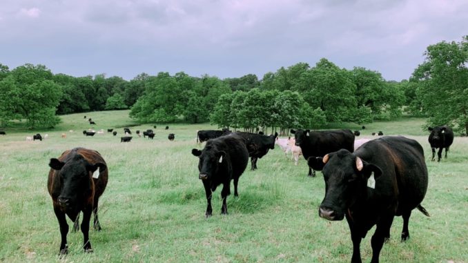
According to the Mesonet, Oklahoma received some much-needed rain in late April (2023). With the moderate temperatures and high humidity, the environment is perfect for the proliferation of gastrointestinal nematodes (GIN) which are commonly called “worms.” Cattle can be infected with a variety of GIN. Most do not cause issues unless husbandry practices are poor. However certain GIN have been associated with disease. The most pathological GIN in cattle is Ostertagia ostertagi. Cooperia species and Haemonchus species are two that have been implicated with production issues. Control of these parasites is constantly changing due to environment, anthelmintic (dewormer) resistance, and consumer preference. Cattle producers should develop a plan to manage these parasites.
In order for GIN to complete their life cycle, certain environmental conditions must exist. The development stage begins with passing of the egg in the feces of the animal. If the egg is to hatch, the temperature must be warm and the humidity needs to be close to 100%. Ideal temperature ranges from 70⁰ to 80⁰ Fahrenheit (F), but any temperature above 45⁰ F will allow for development. Temperatures above 85⁰ F or below 45⁰ F will begin to hamper development. Humidity needs to be 80% or higher.
Once the egg hatches, the larva goes through a couple of molts to reach the infective stage which is the third stage larva (L3). L3 must have moisture to free itself from the fecal pat. Once free, it rides a wave of water on to a blade of forage. Once ingested, this begins the prepatent or pre-adult stage. Two molts take place during this stage (L3 to L4 and L4 to L5). If conditions are not favorable for survivability of offspring, L4 will go into an arrested development stage (hypobiosis) for a period of time. The patent or adult stage is the mature breeding adult.
Once inside the body, the parasite will migrate to certain locations in the digestive tract. For example, O. ostertagi develop in the gastric gland in the abomasum. H. placei and H. contortus will migrate to the abomasum. Cooperia species will live in the small intestine. A few like Trichuris (whipworms) are found in the large intestine.
Clinical signs of parasitism vary according to the species of parasite, burden, and site of attachment. Severe disease, which is referred to as parasitic gastroenteritis (PGE), with internal parasites is unusual with today’s control methods. Clinical signs of PGE are lack of appetite, weight loss, weakness, diarrhea, submandibular edema (bottle jaw), and death. However, most parasite infection are subclinical which means producers do not see clinical signs of disease. In subclinical infections, the parasite causes production issues such as poor weight gain in young cattle, reduced milk production, and lower pregnancy rates.
Producers should be monitoring their herds for parasites throughout the year but especially in the spring when conditions are ideal for infection. A fecal egg count (FEC) is a good way of accessing parasite burdens. Livestock producers need to gather fecal samples from their herd periodically. The samples should be sent to their veterinarian or a veterinary diagnostic lab. Different techniques are used to access the number of eggs per gram of feces. Based on the counts, the producer will learn the parasite burden of the herd. Producers can use this information to develop a treatment plan.
In the past, GIN control was simple. Cattle were routinely dewormed. Unfortunately, anthelmintic resistance has complicated parasite control. Now proper nutrition, grazing management, a general understanding of how weather influences parasites, biosecurity, refugia, anthelmintic efficiency, and the judicious use of anthelmintics are important in designing an effective parasite management program. All of these considerations need to be discussed in detail with a producer’s veterinarian when developing a plan for their operation.
Cattle producers need to understand that parasites cannot be eliminated. They must be managed with a variety of control methods. Designing a parasite management plan requires producers to gain a general understanding of life cycle of the parasite as well as the environmental needs of the parasite. Producers should use this information as well as consult with their veterinarian for a plan to manage GIN. For more information about GIN, producers should talk with their veterinarian and/or with their local Oklahoma State University Cooperative Extension Agriculture Educator.
References
Charlier, J., Höglund, J., Morgan, E. R., Geldhof, P., Vercruysse, J., & Claerebout, E. (2020). Biology and Epidemiology of Gastrointestinal Nematodes in Cattle. The Veterinary clinics of North America. Food animal practice, 36(1), 1–15.
Navarre C. B. (2020). Epidemiology and Control of Gastrointestinal Nematodes of Cattle in Southern Climates. The Veterinary clinics of North America. Food animal practice, 36(1), 45–57.
Urquhart, G. M., Armour, J., Duncan, J. L., Dunn, A. M., & Jennings, F. W. (1987). In G. M. Urquhart (Ed). Veterinary Helminthology. Veterinary Parasitology (1st ed., pp 3-33). Longman Scientific & Technical.
-
 Country Lifestyle7 years ago
July 2017 Profile: J.W. Hart
-

 Outdoors6 years ago
Grazing Oklahoma: Honey Locust
-

 Country Lifestyle3 years ago
The Two Sides of Colten Jesse
-
 Outdoors4 years ago
Pecan Production Information: Online Resources for Growers
-
 Equine7 years ago
Umbilical Hernia
-

 Attractions7 years ago
48 Hours in Atoka Remembered
-
 Farm & Ranch6 years ago
Hackberry (Celtis spp.)
-
 Outdoors3 years ago
Suzy Landess: Conservation carries history into the future

