
Farm & Ranch
Being in a Veterinarian Family

By Barry and Lauren Whitworth
The holiday season will soon be upon us. I hope that this time of year is full of happy memories spent with people you love and hold dear. As all of my children but one have left the nest, the holidays are a special time when I tend to reflect on memories of past experiences with my kids. Although I look back on our holiday memories and experiences with much happiness, my most treasured moments with my children are when they were working with me side by side.
Being the child of a veterinarian might be a curse or a blessing, depending on how you look at it. My kids had a unique “growing up” experience and were used to having plenty animals around. Even to this day when my oldest daughter Lauren sees a new puppy, the first words out of her mouth are, “I want one!” During her college days, when she would spend the summers working in my practice, she would say the same thing about the little “cute” white baby pigs at some of the farms that I would visit on calls. She quickly changed her mind when I finally showed her the sows!
Like most other jobs, I inevitably ended up taking some of my work home with me. My kids never quite knew what they might find inside of our house. On a recent visit home, Lauren walked in to discover that a newly born bottle-fed lamb had taken up residence in our family room.
She smiled and said, “A lamb in the living room; it is good to be home.”
Aside from farm animals in the house, the Whitworth home has some other unique quirks. One holiday tradition that my children were accustom to was the practice of Christmas morning kennel duty. My children were awaken Christmas morning to the words, “Christmas can begin as soon as the dogs are walked and cages are cleaned.”
As are most, my kids were extremely curious about the world around them and ask lots of questions while working with me. Veterinarian kid questions may seem a little bit out of the ordinary for most parents. The questions tended to be about how much blood an animal can lose and still live or about the fairly foul odors that accompany veterinary practice. My son Walker was particularly offended by those smells.
Once on a highly distressing farm call, Walker was holding the tail of a steer with an infected scrotum that both smelled horrid and had a large amount of purulent material. As I proceeded, the smell became too much for him and he began to gag. I laughed and instructed him to “toughen up and hold on to the tail.”
Pale faced, he replied, “You may be used to this, but I am not.”
Life of a veterinarian is messy, but mess is not always a bad thing. Some of my favorite memories are those spent performing C-sections with the help of the whole family. I am not sure why dogs with dystocia tend to occur after hours when help is scarce, but they do. Being the sole veterinarian in my practice, I relied on my family to assist in these late-night cases.
When the distressed patient would arrive, my wife and I would get everything ready for surgery. Then just before administering the anesthesia, Mom would make a call to the house for reinforcements. I am not for sure why my teenage children were tone deaf to the landline phone ringing, but they were. After several rings, my wife would hang up and call one of their cell phones. An almost immediate answer would follow. Mom then gave the instructions, “It is puppy rubbing time!” Reluctantly my teenage children would pile out of bed and file through the clinic doors.
Once the surgery began, my helpers took their positions. Mom would be waiting like a big league baseball player preparing to catch a fly ball. Her hands were covered with a towel ready for me to hand her that first pup. She then handed off to one of the kids who began vigorously rubbing the life into these little dogs.
The procedure was repeated until all the puppies were born. In my many emergency C-sections, it never failed that some puppies were reluctant to wake up and join the world. We usually assigned the stubborn puppies to my middle daughter Madison. She was always up for the challenge and worked tirelessly to stimulate their breathing. We surmised that she was gifted at irritating her brother and sister, so she should be able to irritate the pup to wake up.
After all the puppies had nice red noses, were crying, and mama dog was being stitched up, my wife would dismiss the troops with instructions to get to bed because it was late. As my assistants headed home, most of my clients would comment on what hard workers my children were and how proud I must be of them. I would simply smile and say, “Yes, I am very proud of them.”
I hope that as you go through life that you are making memories with your children. In my experience, they grow up and leave home. Do not wait for special occasions to spend time and share life with your kids. Every day is a good time to make a memory. My family and I wish you and your family a Merry Christmas. May God bless you with good health and happiness in the New Year.

By Savannah Magoteaux, Editor
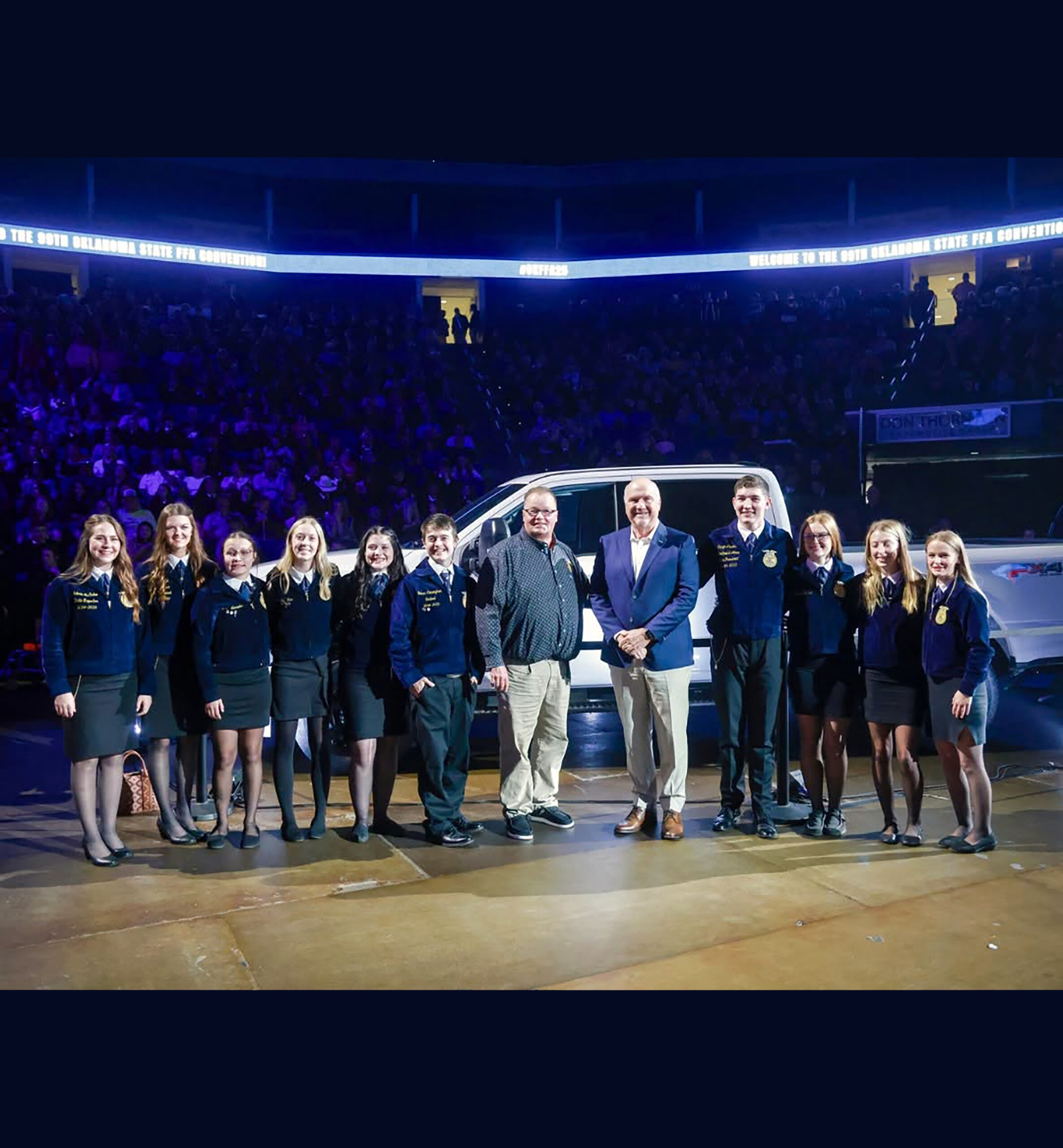
Bluejacket, Oklahoma, is the kind of town where almost everyone knows everyone else. With a population of about 300 and a K-12 enrollment of about 170 students, it is not where most people would expect to find an FFA chapter sending students across the country, much less across the Atlantic Ocean.
But Bluejacket FFA Advisor Shawn Martin has never believed the size of a student’s hometown should determine the size of his or her opportunities.
Under Martin’s leadership, Bluejacket FFA has grown from about 33 members to between 65 and 70 each year. Students have traveled to National FFA Convention, toured agriculture in Ireland, visited the John Deere factory in Iowa, earned National Three-Star Chapter recognition and been selected for a national chapter exchange program. At home, they weld, build projects, grow produce, sell at farmers markets and help provide fresh food to people in their community.
That list would be impressive anywhere. It stands out even more at a school where a graduating class may number only a dozen students.
Yet Martin doesn’t talk about any of it as though he built the program alone. He keeps coming back to the same point: the students have to buy in.
“The most important thing is getting the buy-in to kids and getting them to want to go and see and do,” he said. “I’m very proud of them. We have lots of kids participating and helping and making these things happen.”
That belief in giving students ownership may be one of the biggest reasons Bluejacket FFA has grown into the program it is today.
Coming home
Martin understands small-town life because Bluejacket has always been home.
He was born and raised in the area, attended Bluejacket schools and was deeply involved in FFA himself. He served two years as chapter president, earned his State FFA Degree and graduated in 1987.
“I just loved everything about the FFA,” he said. “We had a great program there when I was a kid too.”
He considered teaching agriculture early on, but his career went another direction. After attending college for a time, Martin saw the earning potential in pipe welding and went to work in the trade.
He spent about 22 years as a pipe welder, traveling across the country. The work paid well, but it kept him away from home for much of his life. At one point, Martin estimates, he was on the road around 330 days a year.
Eventually, the trade-off became harder to accept. The birth of his grandson brought the decision into focus.
“I came home and I saw him, and I’m like, ‘I’m not doing this anymore. I’m not going to miss these grandkids growing up like I did with the kids growing up,’” Martin recalled.
He couldn’t walk away from a successful welding career overnight. His family was used to the income, and Martin knew he had to prepare financially before making the change. He spent several years putting himself in a position to come home and go back to college.
In the end, the decision came down to what he wanted the next part of his life to look like.
“At some point it’s not worth it anymore, and the money’s not what it’s all about,” he said. “You need some kind of personal happiness and something that gives you meaning. Sitting alone in a hotel room 800 miles away from your own house and your own family, it gets old.”
Martin retired from welding around 2011 and finished his education degree by 2015. Soon afterward, an agriculture teaching position opened at Bluejacket. It was his first full-time teaching job, and his farm was only three miles away.
The boy who once wore the Bluejacket FFA jacket had come home to lead the same program.
Building pride
When Martin took over, the chapter had about 33 members. He felt the program had grown complacent and wasn’t taking advantage of many of the activities and opportunities FFA offers.
Before chasing awards or adding trips, Martin wanted the students to be proud of their own program.
“My first thing when I came back, I wanted to put that pride in that ag program,” he said.
He started with the classroom. Martin remembered walking into a plain white room with little sense of identity or ownership. He remodeled it with rusted tin and gave it a farmhouse feel.
“This is your place, and you guys take pride in it,” he wanted the students to understand.
Students started wearing Bluejacket FFA apparel and joining more activities. Slowly, Martin saw them take ownership.
“We started getting the kids to buy in, and they really did,” he said.
Membership grew. Today, 65 to 70 students are involved each year, most of the students eligible to participate.
A major turning point came when a group of younger members set their sights on National Three-Star Chapter recognition. By Martin’s third year, they had earned it.
“Those kids had set that goal, and like, ‘This is what we want to do,’ and that was a turning point for the chapter right there,” he said.
For Martin, the recognition confirmed that the hours going into the program were paying off.
“It was overwhelming because you put all these hours in, and you’re like, ‘Gosh, I don’t know if we’re getting where we need to get,’” he said. “But when that happened, I’m like, ‘Okay, we’re on the right track, and we’re doing good things.’”
The students traveled to Indiana and crossed the stage at National FFA Convention.
“It blew me away,” Martin said. “I was so excited. The kids were so excited. Got to go to Indiana and cross that national stage and hear our chapter name mentioned.”
Bluejacket is pursuing National Three-Star recognition again, and Martin doesn’t do the work behind the scenes for his students. The application requires extensive documentation of chapter activities and impact, and he expects his officers and members to handle it.
“I don’t sit and fill out their applications and all that stuff for them,” he said. “I help where they need help, but they have to buy into it.”
Seeing the world
That approach carries over into other opportunities, too. When Martin talks about what he wants to give his students, travel comes up quickly.
He enjoys traveling himself and sees great value in taking young people beyond the places they already know. Some Bluejacket students, he said, had not traveled far before joining the program.
“I personally love to travel too, and I’ve kind of encouraged the kids to do that,” he said.
A few years ago, Bluejacket FFA took 10 students to Ireland for an agricultural tour of the countryside.
More recently, Martin and his students asked how they could create a trip for the entire chapter. Through a separate fundraising effort, they took students to Waterloo, Iowa, where they toured the John Deere factory that builds the company’s large 9 Series tractors. The trip also included the Field of Dreams and time at water parks.
For Martin, mixing agriculture with experiences students enjoy is part of the point. He wants them to learn, and he also wants them to discover how big the world is outside their hometown.
“Getting them out of state and showing them how much fun traveling is and what cool places there are to see,” he said.
The trips do more than make memories. Martin hopes seeing different places, careers and industries will change the way students think about their futures.
“If we can take them places and show them these things, maybe you might decide, ‘Hey, I want to go to college,’ or, ‘Hey, I want to go get me a career of some kind, even in the trades, where I can make good money,’” he said.
Martin doesn’t discourage students from staying close to home. He wants staying home to be a choice rather than the result of never knowing other options existed.
“Staying local is great if that’s what you want to do,” he said.
He also talks candidly with students about earning a living and building a stable future.
“I know it’s not all about money, but making a good living takes a lot of pressure off of you in your life,” Martin said.
His own career gives him practical experience when those conversations turn to skilled trades. Martin teaches welding at Bluejacket, and every student gets at least some time behind the hood. He can also tell students what a welding career looks like, including the income and the sacrifices that come with traveling work.
Students tend to focus on the paycheck.
“The first thing they want to know is how much money you made, and then that’s it,” Martin joked. “They don’t hear anything after that.”
Still, his advice stays the same. College is not the only path to success, but he wants students to pursue some kind of education or career training after graduation.
The students do the work
The same philosophy of preparation and responsibility runs through the chapter’s activities.
When Bluejacket applied for a national Chapter Connect program, Martin didn’t fill out the application. The students did. He told them about the opportunity and asked whether they wanted to pursue it.
“‘What, you guys want to do it? Fill it out,’” he recalled telling them.
They wrote the application, brought it to him to proofread and submitted it. Bluejacket was selected.
The program pairs FFA chapters from different parts of the country, and Bluejacket was matched with a chapter from an urban area of Virginia. The students will work together online on projects throughout the year, with plans for an exchange that lets each group see the other’s community and agriculture.
When the Virginia students come to Oklahoma, Martin plans to show them what agriculture looks like around Bluejacket. One stop will be the Layton family ranch west of Welch, where students can see horses, cattle and a working ranch and learn about rodeo culture from Landry Layton.
For Bluejacket students, the exchange offers another chance to see a very different part of the country. It is exactly the kind of opportunity Martin wants them to pursue, but he also wants them to understand that opportunities take effort before they ever become available.
“They have to buy into it,” he said.
Bluejacket students compete, and Martin is proud when they succeed.
“My kids get a lot of plaques, and they do great,” he said.
He also wants agricultural education to stay connected to agriculture itself. At Bluejacket, students weld. They build chicken coops, chicken tractors, outdoor furniture and other projects. Before building, Martin makes students draw plans and figure out what materials they need.
“I want everybody to build one project,” he said. “The first thing you do is draw me a set of plans. Then, what materials do you need before ordering?”
The goal is to teach students to think a problem through from beginning to end.
“You may be surprised one day you want to build something at your house, and you could do it,” he tells them.
Martin knows not every student will become a welder or work in production agriculture, but he believes the confidence that comes from learning to build, repair, plan and solve problems pays off in any career.
The same goes for public speaking. Martin remembers one student who told him in eighth grade that she would never give a speech.
“She told me, ‘I’m never going to give a speech,’” he said.
By her senior year, she was almost unrecognizable from that reluctant eighth grader.
“That girl could walk in front of 300 people and control that room and run it and knows how to speak, and just became so good at it,” Martin said. “That’s the stuff I really love to see.”
Those transformations are one reason he resists defining the program by contests and awards alone.
“It’s not all a contest,” Martin said. “They have their place, and they are important. But it’s not all there is to agriculture.”
He sometimes wonders whether agricultural education leans so heavily toward contests that students miss basic hands-on skills.
“Are we forgetting our roots, and are we forgetting that we still need to know these fundamentals of agriculture, hands-on?” he said.
At Bluejacket, Martin is determined that the answer be no.
Growing food, giving it away
You can see that commitment on the school’s 10-acre property. What started as a small garden several years ago has grown into a large outdoor garden, an orchard and a 40-by-120-foot high tunnel. Students help plant, install watering systems, harvest produce and learn what it takes to grow food. They also grow hay on part of the property.
The produce operation now supplies tomatoes, cucumbers and other vegetables to the school and brings in income for the FFA chapter through farmers markets. Martin estimates the chapter earned around $10,000 to $11,000 from produce sales over the summer.
The chapter has also partnered with the Cherokee Nation, accepting its farmers market benefits and helping bring fresh produce to tribal citizens in the region. At times, the students take produce to Bernice so it is closer to people in tribal food distribution programs.
Not everything the students grow gets sold. When there is extra produce, Martin said, the chapter shares it with about 11 families or individuals, including elderly and homebound people and others who may need help.
Most people never see that part of the program.
“That’s something you’ll never see on the Facebook page,” Martin said. “We don’t talk about it.”
That is intentional. Martin wants his students to learn that service doesn’t need recognition.
“When you do something nice, don’t take pictures of it and put it on Facebook,” he tells them. “Just do it to be doing it.”
For all the national recognition, travel and contest success Bluejacket FFA has earned, that lesson may say more about what Martin wants the program to accomplish than any plaque on the wall.
“We give a lot of it away to families all through the summer months so they can have fresh produce too,” he said. “I’m trying to teach them to be good and to help people when you can, because I think that’s important too.”
It is another kind of preparation for life, one you can’t measure with a contest score or list on an application.
“We’re a small school, but we’re trying to do good things and grow the program and teach the kids the right way,” Martin said. “Be a contributor in the community too.”
Martin once left Bluejacket to make his living hundreds of miles from home. Coming back gave him the meaningful second career he was looking for. Now his work centers on making sure the young people growing up in that same small community see their hometown as a starting point rather than a limit.
They may go to college. They may learn a trade. They may stay in Bluejacket or travel far beyond it. Martin sees his job as making sure they have seen enough, learned enough and done enough to know they have choices.
And more and more, his students are taking responsibility for creating those opportunities themselves.
By Barry Whitworth, DVM
Recently, I accompanied a County Extension Ag Educator on a farm visit about concerns of blue-green algae toxicity. A few days later, another County Extension Educator emailed me about a cattle producer with the same concerns. With hot, dry weather across parts of Oklahoma, livestock producers should be watching their ponds closely for blue-green algae.
Blue-green algae is not an algae at all. It is a bacterium, called a cyanobacterium. The most common species in the Midwest are Microcystis, Oscillatoria and Anabaena. The bacterium lives in most bodies of water, but it becomes a problem during periods of rapid growth fueled by high nitrogen and phosphorus levels and warm, sunny weather. Most cyanobacteria problems occur in summer and fall, but high toxin release can happen any time of year. Oklahoma has recorded cases as early as March.
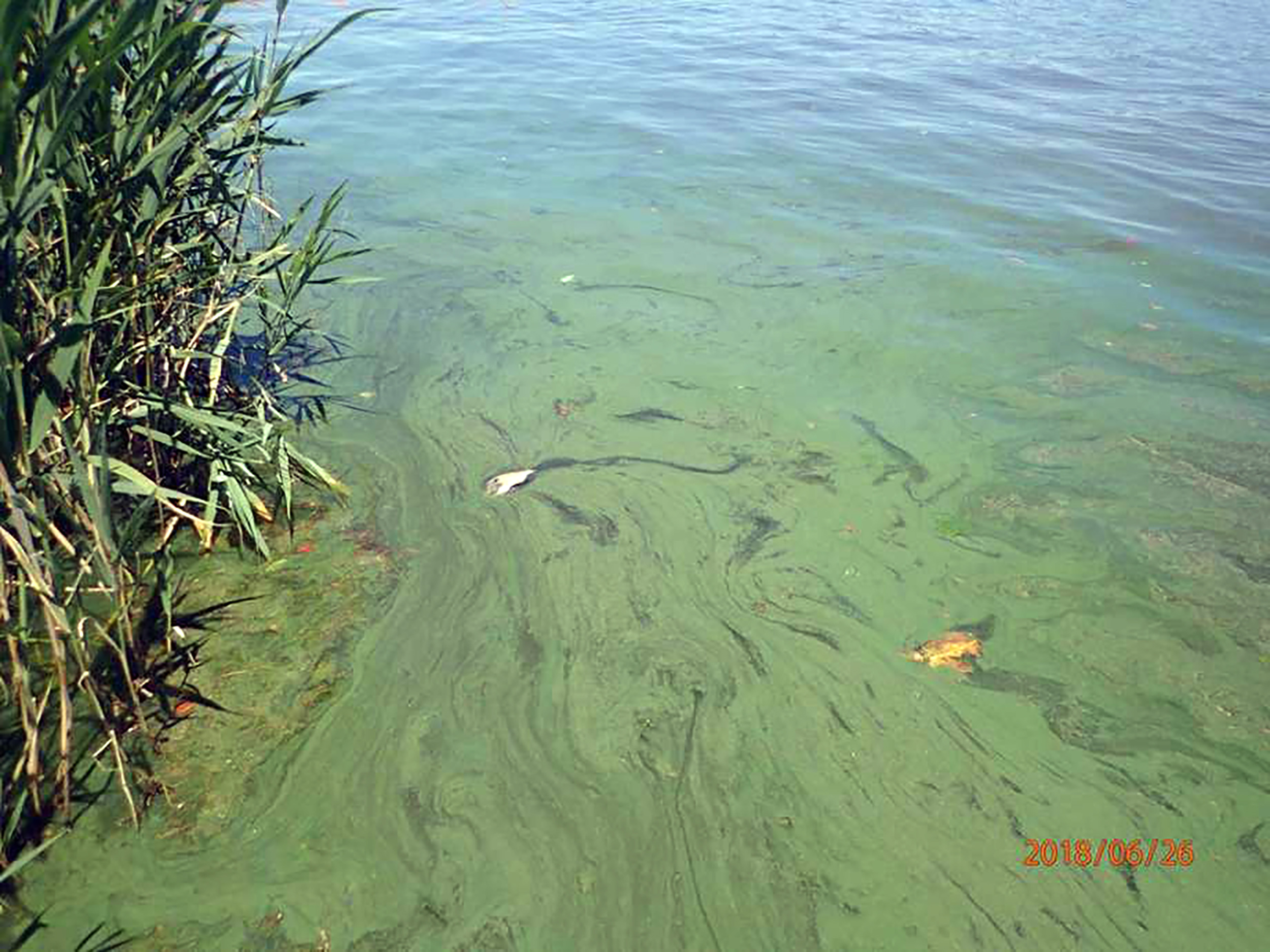
When the bacteria overgrow, they die and float to the top, forming a scum on the water. The scum usually looks blue-green, but its color can vary. Wind can push the scum around the pond and concentrate toxic levels of dead bacteria in certain areas. Rain or wind can break up the scum and lower the risk, but not always.
All livestock, pets, wild animals and humans are susceptible to blue-green algae toxicity. The amount of water needed to cause toxicity depends on the species of animal, the concentration of toxins in the water and how much water the animal drinks. One quart of highly concentrated water can kill a cow.
Most producers don’t recognize a blue-green algae problem until they find dead livestock in or near a pond. Most cattle that drink contaminated water die, though producers occasionally find sick cattle. Clinical signs depend on which toxin the animal ingested. Blue-green algae produce two types: neurotoxins, which affect the nervous system, and hepatotoxins, which affect the liver. Cattle affected by a neurotoxin show muscle tremors, reluctance to move and breathing problems early on, followed by convulsions and death. Cattle that drink water containing liver toxins develop weakness, pale mucous membranes, gastroenteritis, nervous signs and death. Animals that survive lose weight and become poor doers. Survivors may also develop photosensitization, which makes light-colored skin prone to sunburn.
Veterinarians typically base a diagnosis on exposure to blue-green algae along with clinical signs or sudden death. If you suspect blue-green algae killed your cattle, collect a water sample right away from where the algae is heaviest. Wind can disperse the toxin, so speed matters. Then call a veterinarian to perform a necropsy, which will rule out other causes of death. The veterinarian will likely take tissue samples for testing and may examine rumen contents for blue-green algae.
The water sample will need to go to a laboratory for analysis. For guidance on necropsy or water sampling, contact the Oklahoma Animal Disease Diagnostic Laboratory at (405) 744-6623 or visit https://oaddl.okstate.edu/.
There is no known antidote, and treatment rarely works. Producers need to focus instead on the conditions that favor blue-green algae. Algae bloom in sunny, warm weather, and the worst blooms occur in ponds that catch runoff high in nutrients. Inspect ponds any time these conditions are present, and be ready to provide another water source in a crisis.
Blue-green algae toxicity is not a new problem for Oklahoma livestock. When the weather favors algae growth, watch your ponds for blue-green scum on the water, and act quickly if you find it. Oklahoma State University Extension offers an excellent fact sheet on blue-green algae and how to manage it at https://extension.okstate.edu/fact-sheets/toxic-blue-green-algal-blooms. If you have questions about blue-green algae, contact your local veterinarian or your Oklahoma State University County Extension Agriculture Educator.
References
Meehan, M. A., Galbreath, J., & Mostrom, M. (2026). Cyanobacteria (blue-green algae) poisoning [Fact sheet]. North Dakota State University Extension.
Morgan, S. E. (2011). Water quality for cattle. Veterinary Clinics of North America: Food Animal Practice, 27(2), 285.
Newcomer, B. W. (2022). Toxicologic insults to the bovine liver. Veterinary Clinics of North America: Food Animal Practice, 38(3), 421–432.
Osweiler, G. D., Carson, T. L., Buck, W. B., & Van Gelder, G. A. (1985). Toxic blue-green algae. In Clinical and Diagnostic Veterinary Toxicology (3rd ed., pp. 451–452). Kendall/Hunt.
By Tony Dean, tonydean.tx1@gmail.com
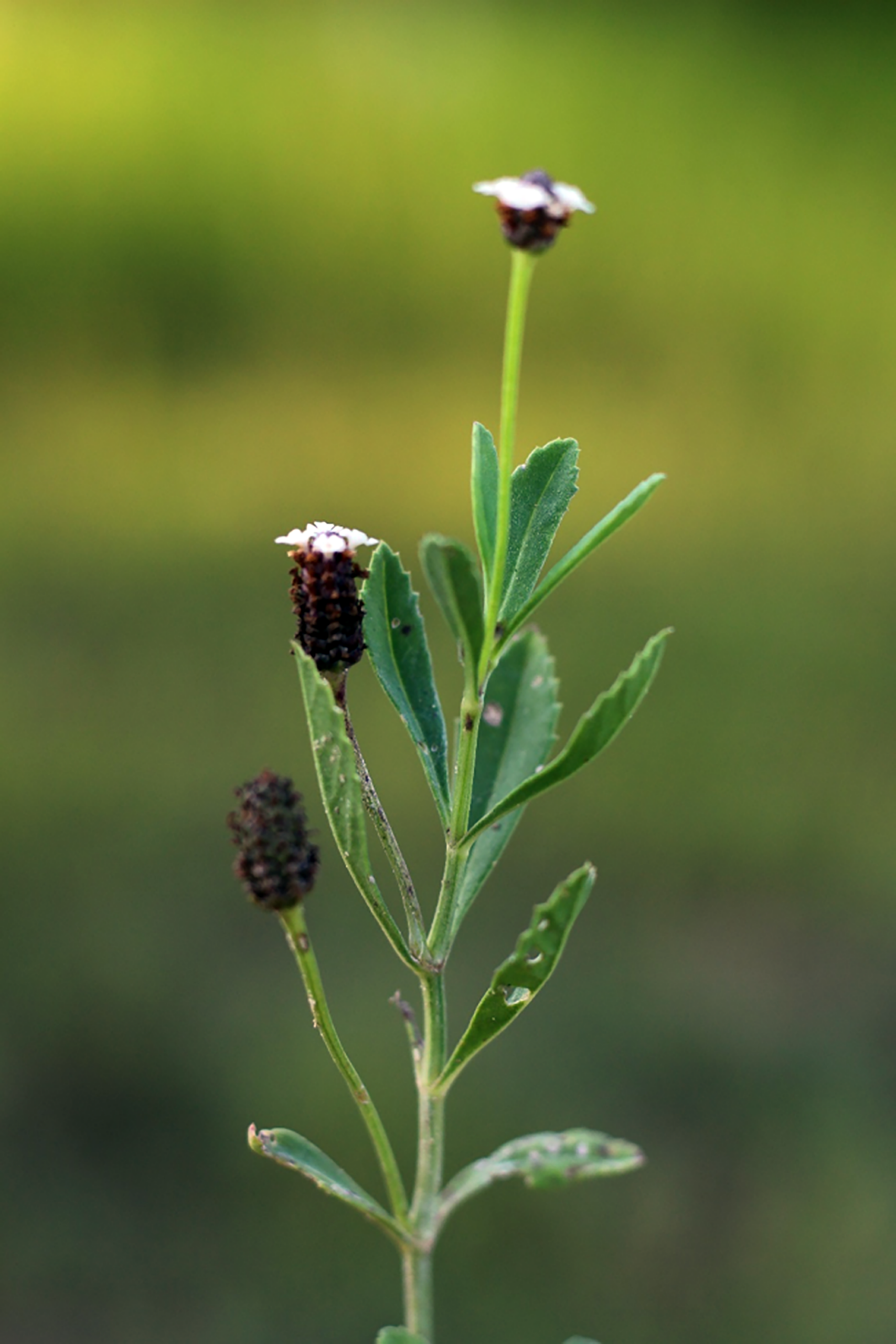
Frog fruit is a native, low-growing perennial forb adapted to most of the eastern U.S., but few grazers know it well. It contributes more to our plant communities than we realize. It generally grows no more than a few inches tall and roots at the nodes. It can form dense mats several feet across and develop runners up to 3 feet long.
The leaves are thick, one to two inches long and 3/8 inch wide, and toothed from the middle to the tip. Tiny white flowers form a ring around a flower head, and the head then elongates into a cylinder an inch long or longer. Frog fruit has a four-angled stem.
Frog fruit can grow almost anywhere in Texas and on many soil types. It can grow in drier soils and likes good drainage, but it needs additional moisture to survive the hottest months. You can find it almost anywhere in a pasture, though it often grows in depressions and near water sources.
“Frog fruit is used as forage by livestock and deer. I recall an instance when I was moving cattle from one pasture to the next on a Jack County ranch. As I opened the gate, I stood by a beautiful green plant of switchgrass which I expected to be immediately grazed down. The cattle walked right by the switchgrass and stuck their heads in a small patch of frog fruit a few feet away,” Dean said.
Frog fruit is gaining popularity as a ground cover for homes and gardens for these reasons:
- Frog fruit grows quickly, maybe too quickly. If it gets out of bounds, you can always trim it back.
- Frog fruit is low maintenance. Once you get it established with adequate watering, it can survive with little care.
- Frog fruit has a long bloom period, from April to October.
- It is a host plant for several butterfly species and other insects.





-
 Country Lifestyle2 years ago
July 2017 Profile: J.W. Hart
-
 Farm & Ranch2 years ago
From Plow to Plentiful: The Most Important Inventions in Agricultural History
-
 Attractions9 years ago
48 Hours in Atoka Remembered
-
 Equine9 years ago
Umbilical Hernia
-
 Country Lifestyle5 years ago
The Two Sides of Colten Jesse
-
 Outdoors9 years ago
Grazing Oklahoma: Honey Locust
-
 Farm & Ranch8 years ago
Hackberry (Celtis spp.)
-
 Attractions3 months ago
Oklahoma Ghost Towns – Navajoe