
Farm & Ranch
Theileria orientalis Genotype Ikeda: an Emerging Cattle Disease?

Theileria orientalis genotype Ikeda is a hemoprotozoan. A hemoprotozoan affects red blood cells and leukocytes. T. orientalis genotype Ikeda has been associated with severe bovine disease in Japan, New Zealand and Australia (Watts, Playford, & Hickey, 2016). In August of 2017, the protozoan was found associated with illness and death in cattle at a ranch in Virginia (Oakes et al., 2019). These cattle displayed clinical signs of weakness and anemia. A preliminary diagnosis of anaplasmosis was given. Blood samples taken from the animals were tested for Anaplasma, Babesia and Leptospira. Results from the test revealed the cattle were all negative; however, a blood protozoan was detected. This protozoan was identified as T. orientalis genotype Ikeda (Oakes et. al, 2019). Since this original herd outbreak, other herds of cattle in Virginia and West Virginia have been found to be infected with the organism.
In Australia, the Asian longhorn tick (Haemophysalis longicornis) has been identified as a possible vector of T. orientalis genotype Ikeda (Hammer, Emery, Bogema, & Jenkins, 2015). In 2017, the United States Department of Agriculture’s National Veterinary Services Laboratories confirmed the presence of Haemaphysalis longicornis, which is commonly referred to as the Asian longhorn tick or bush tick. In an effort to understand how the tick got to the United States, USDA officials discovered the tick had been found in West Virginia in 2010. The tick has been confirmed to be in Arkansas, Connecticut, Delaware, Kentucky, Maryland, New Jersey, New York, North Carolina, Pennsylvania, Tennessee, Virginia, and West Virginia (USDA, 2020). Some evidence exists for other possible insect vectors for T. orientalis genotype Ikeda. Needle transfer is another possible route of transmission of the organism (Watts, Playford, & Hickey, 2016).
If the Asian longhorn tick is ever found in Oklahoma, Dr. Justin Talley, Oklahoma State University Extension livestock entomologist, believes the tick will live east of I-35. The tick will likely be found in vegetation that is high in humidity in areas such as woods, brush or tall grass. Ticks are usually found where large numbers of wildlife congregate such as a deer trail. Dr. Talley and Dr. Bruce Noden have written an excellent fact sheet about the longhorn tick. The fact sheet can be found at http://entoplp.okstate.edu/pddl/2018/PA%2017-16.pdf.
Cattle infected and sick with T. orientalis genotype Ikeda will have clinical signs of fever, weakness, anorexic, and exercise intolerance. If cattle are forced to move, they may stagger and gasp for air. If stressed too much, the cattle may collapse and die. When examining cattle, the gums, eyes or vaginal mucosa may appear white or yellow in color. Reproductive losses including stillbirths, and late term abortions may be seen as well as reduction in milk production (Spickler, 2019). Since Anaplasma marginale and T. orientalis genotype Ikeda both display similar clinical signs, one difference that has been noted in the two diseases is A. marginale infected cattle usually display aggression and T. orientalis genotype Ikeda do not. Still, a laboratory test would have to be performed to differentiate the two diseases.
In other countries, therapies have been developed to treat this organism. Unfortunately, no approved treatments are available in the United States. Also, there are no vaccines for this disease. The best defense to this disease is to control ticks. This usually requires a combination of insecticide treatments and pasture rotation to avoid areas such as woods and brushy areas where ticks live.
Theileria orientalis genotype Ikeda is not likely to arrive in Oklahoma anytime soon and in reality, may never become a major problem in Oklahoma or the United States. However, producers need to keep in mind the natural progression of cattle in the United States is east to west and south to north. Oklahoma is unique in the fact large numbers of cattle move from the southeast United States to graze on grass and wheat in our state. From there, the cattle go to the feedyards. The tick and organism could easily be transported to Oklahoma on a load of stockers headed for grazing or to a feedlot. If a producer would like more information on T. orientalis genotype Ikeda, they should contact their local veterinarian or Oklahoma State University Extension Educator or visit the Center for Disease Control and Prevention at https://wwwnc.cdc.gov/eid/article/25/9/19-0088_article.
References
Hammer, J. F., Emery, D., Bogema, D. R., & Jenkins, C. (2015). Detection of Theileria orientalis genotypes in Haemaphysalis longicornis Ticks from Southern Australia. Parasites & vectors, 8, 229.
Oakes, V. J., Yabsley, M. J., Schwartz, D., LeRoith, T., Bissett, C., Broaddus, C., Schlater, J. L., Todd, S. M., Boes, K. M., Brookhart, M., & Lahmers, K. K. (2019). Theileria orientalis Ikeda Genotype in Cattle, Virginia, USA. Emerging infectious diseases, 25(9), 1653–1659.
Spickler, Anna Rovid. (2019).Theileriosis. Retrieved from http://www.iastate.edu/DiseaseUbfi/factsheets.php.
Watts, J. G., Playford, M. C., Hickey, K.L. (2016). Theileria orientalis: A Review, New
Zealand Veterinary Journal, 64:1, 3-9.
Read more great stories in the April 2020 issue of Oklahoma Farm & Ranch.
Barry Whitworth, DVM | Area Food/Animal Quality and Health Specialist for Eastern Oklahoma
*Article originally printed in the October 2022 issue of Oklahoma Farm & Ranch.
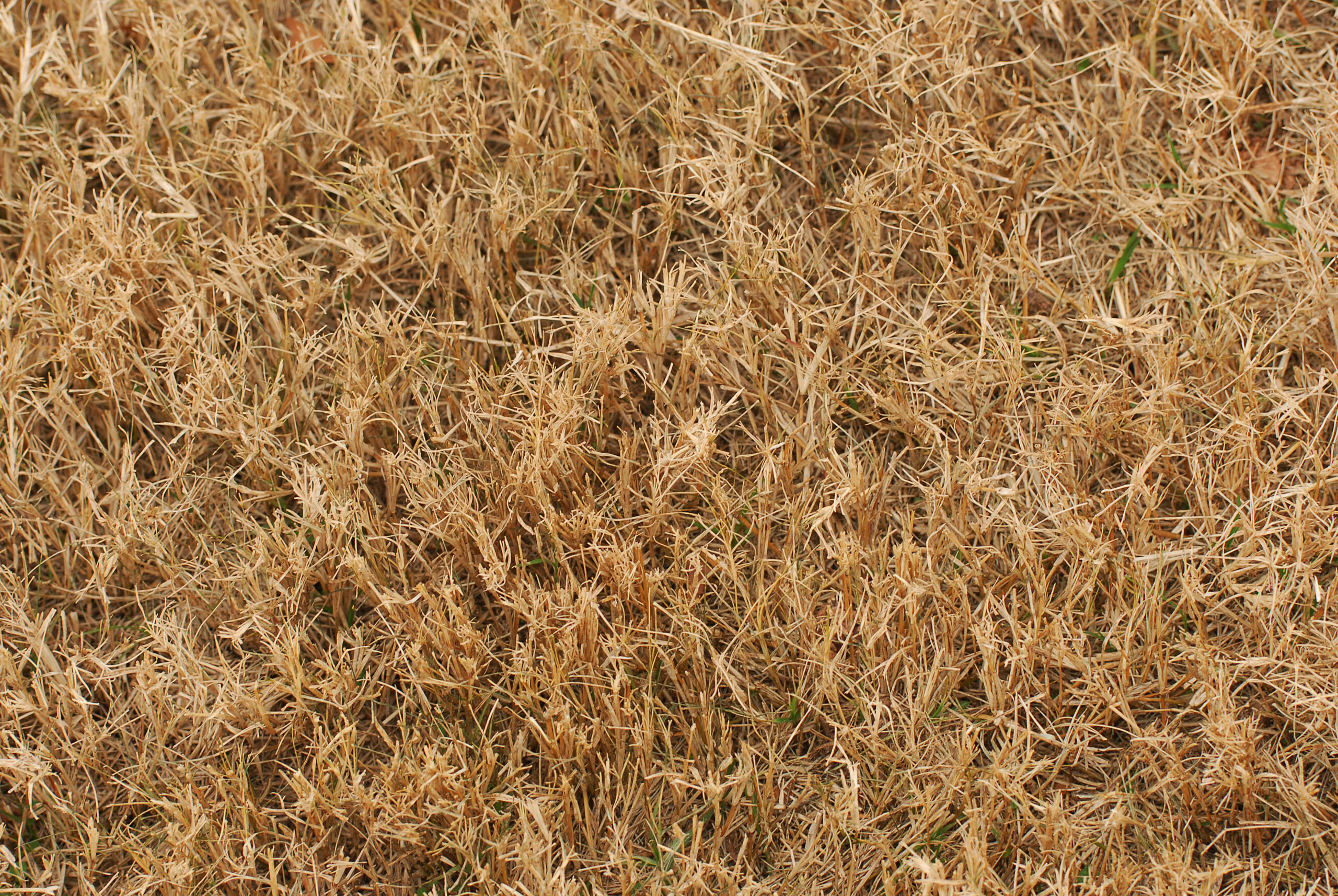
Since most of Oklahoma experienced drought conditions and with fall fast approaching, producers with fescue pastures should closely observe their livestock for any signs of fescue toxicity. According to Mike Trammel, Pottawatomie County Ag Educator and Muti-County Agronomist, fescue toxins (ergot alkaloids) tend to increase in Kentucky-31 tall fescue pastures in the fall. Some reports indicate more problems with fescue toxins following a summer drought and limited fall rains. All of this may put Oklahoma cattle at a greater risk of fescue toxicity.
One issue that cattle experience with fescue toxins is fescue foot. Fescue foot is thought to be caused by ergot alkaloids such as ergovaline. These alkaloids are produced by endophyte fungus (Epichloë coenophiala) which is in tall fescue. Ergovaline has been proven to be a vasoconstrictor which might be responsible for fescue foot and heat intolerance also known as summer slump in cattle. Other issues that may be seen with the ergot fescue toxins are reduced milk production and reproductive issues.
Clinical signs of fescue foot appear within a few days of cattle being turned on to tall fescue pastures or it may take weeks if toxins in the pasture are low. Producers will initially observe cattle with arched back, rough hair coats, and sore feet. These symptoms are more noticeable early in the morning and with cold weather. This is followed by reddening and swelling in the area between the dewclaws and hooves. The lameness usually becomes more severe with time. If no action is taken, gangrene will result in loss of tissues distal to the coronary band and declaws. If the weather remains mild, other signs such as increase respiration rate, increase heart rate, and higher body temperature are more common.
Other causes of lameness in cattle must be differentiated from fescue foot. One simple method that will help differentiate fescue foot from footrot is to check the temperature of the foot. If the foot is cold, this is an indication that the problem is more likely fescue foot.
Since there is not a specific treatment for fescue foot, the condition must be managed. Cattle need to be observed daily for any signs of lameness or stiffness during the first few weeks on fescue pastures. This should be done early in the morning before cattle walk off the stiffness. Producers should pay close attention during cold weather, especially when rain, snow, or ice are present. Any animal showing clinical signs of fescue foot should be removed from the pasture and placed in a clean environment. The animal should be fed a ration with no fescue toxins.
The best but most costly solution to reduce fescue toxicity is to renovate old pastures with new endophyte friendly varieties. If this option is not possible, producers might try interseeding fescue pastures with clovers or other grasses. This should dilute fescue toxins. Nitrogen fertilization may increase ergot alkaloids, so producers should avoid fertilizing fescue pastures with high amounts of nitrogen. Researchers have demonstrated that feeding a supplement while grazing fescue pastures reduces clinical symptoms. Some studies indicate a difference in susceptibility to fescue toxicity in some cattle. Selecting cattle based on genetic tolerance of fescue toxins is an option. (For more information go to www.agbotanica.com/t-snip.aspx)
With large areas in Oklahoma covered with Kentucky-31 fescue pastures, fescue foot as well as other fescue toxicities are not going away any time soon. Livestock producers will need to watch their livestock closely for any signs of fescue toxicity and manage their pastures to keep toxins as low as possible. If producers would like more information on fescue foot, they should consult their veterinarian and/or visit their local Oklahoma State University Cooperative County Extension Agriculture Educator.
By Barry Whitworth, DVM, MPH | Senior Extension Specialist Department of Animal & Food Sciences | Ferguson College of Agriculture | Oklahoma State University
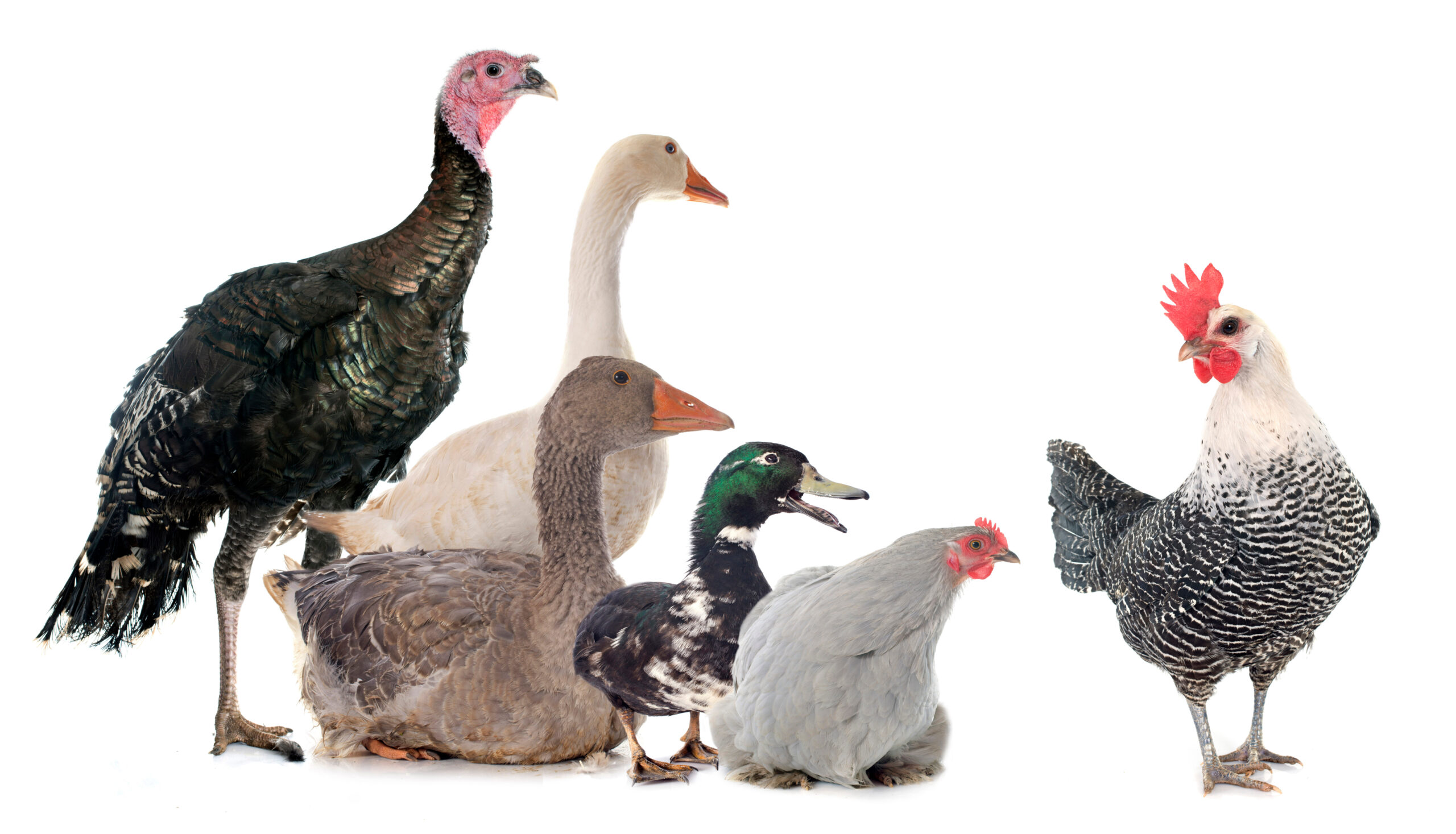
According to the 11th edition of Poultry Diseases, external parasites of poultry are arthropods that live on or in the skin and feathers. Essentially, parasites are freeloaders that live at the expense of the host. Backyard birds are infested with a variety of pests. Ticks, fleas, mites, and lice are some of the most common external parasites found in chickens, turkeys, and ducks. Several of these parasites are bloodsuckers. If not controlled, they can cause weight loss, decreased egg production, unthriftiness, and death in severe cases.
According to a study conducted by Dr. Amy Murillo and associates in California, the most common external parasites in backyard flocks were lice, fleas, and mites. Lice were the most frequently observed parasites, with the chicken body louse (Menacanthus stramineus) found on half of the premises inspected. The fluff louse (Goniocotes gallinae) was found in 35% of operations. The wing louse (Lipeurus caponis) and sticktight flea (Echidnophaga gallinacea) were present in 20% of flocks. Northern fowl mites (Ornithonyssus sylviarum), which are the most common mites found in commercial poultry operations, were detected in only 15% of flocks. However, the survey was conducted in the summer, which may have influenced the low number of northern fowl mites, since they are most active in the winter.
Birds infested with external parasites often become agitated due to skin irritation. They will spend more time preening and scratching. Their feathers may become damaged, and they may appear unhealthy. Birds showing these signs should be examined.
When examining birds for external parasites, producers should focus on the breast, back, head, vent region, and wings. Lice may be found on different parts of the body. They are yellowish in color and lie flat against the skin. Their eggs are typically found attached to the shafts of feathers. The vent area is the primary location to check for mite infestations and may appear “dirty.” Sticktight fleas are usually found embedded in the comb.
Birds should be monitored regularly. When producers are unable to examine all birds, they should focus on the young, the old, and any bird that appears unhealthy. The coop should also be inspected. Producers should examine the bedding, walls, and roosts, with close attention given to crevices and cracks where pests may hide.
Before parasite control can begin, the parasite must be correctly identified. Producers can use books or other publications for this purpose, or they may consult a veterinarian. Contacting the local Oklahoma State University Extension office is also a useful option. An agricultural extension educator may be able to identify the pest or submit samples to the Plant Disease and Insect Diagnostic Laboratory at Oklahoma State University for identification.
Prevention and control of external parasites require an integrated approach. The first line of defense is a strong biosecurity program to prevent parasites from entering the operation. Sanitation is also critical, keeping the coop and surrounding area clean helps prevent infestations.
Maintaining healthy birds is essential in preventing parasite infestations. Producers should focus on proper nutrition and disease prevention as they are key factors in maintaining a healthy flock. A strong immune system can help birds better withstand some external parasites.
Selecting the proper pesticide and using it correctly is essential. Many pests described in this article can be controlled with appropriate pesticides; however, their eggs are not killed, which requires repeated applications to target newly hatched larvae. Producers should read and follow pesticide label directions.
Alternative methods for external parasite control are also available such as providing diatomaceous earth mixed with sand for dust bathing or using sulfur bags to control mites and lice. For more information on these methods, see references below.
Finally, early identification and treatment greatly increase the chances of successful control. If infestations are allowed to become established, control becomes much more difficult.
For more information on external parasites in backyard poultry, producers may visit https://www.veterinaryentomology.org/ or contact their local veterinarian or Oklahoma State University County Agriculture Extension Educator.
References
Arends, J., J. (2003). External parasites and poultry pests. Diseases of Poultry. 11th Edition.
Murillo, A. C., & Mullens, B. A. (2016). Diversity and Prevalence of Ectoparasites on Backyard Chicken Flocks in California. Journal of medical entomology, 53(3), 707–71.
Murillo, A. C., & Mullens, B. A. (2016). Timing Diatomaceous Earth-Filled Dustbox Use for Management of Northern Fowl Mites (Acari: Macronyssidae) in Cage-Free Poultry Systems. Journal of economic entomology, 109(6), 2572–2579.
Murrillo, A.C., Mullens, B.A. (2016). Sulfur Dust Bag: A Novel Technique for Ectoparasite Control in Poultry Systems: Journal of Economic Entomology, 109(5), 2016, 2229-2233.
Barry Whitworth, DVM
Senior Extension Specialist Department of Animal & Food Science Ferguson College of Agriculture
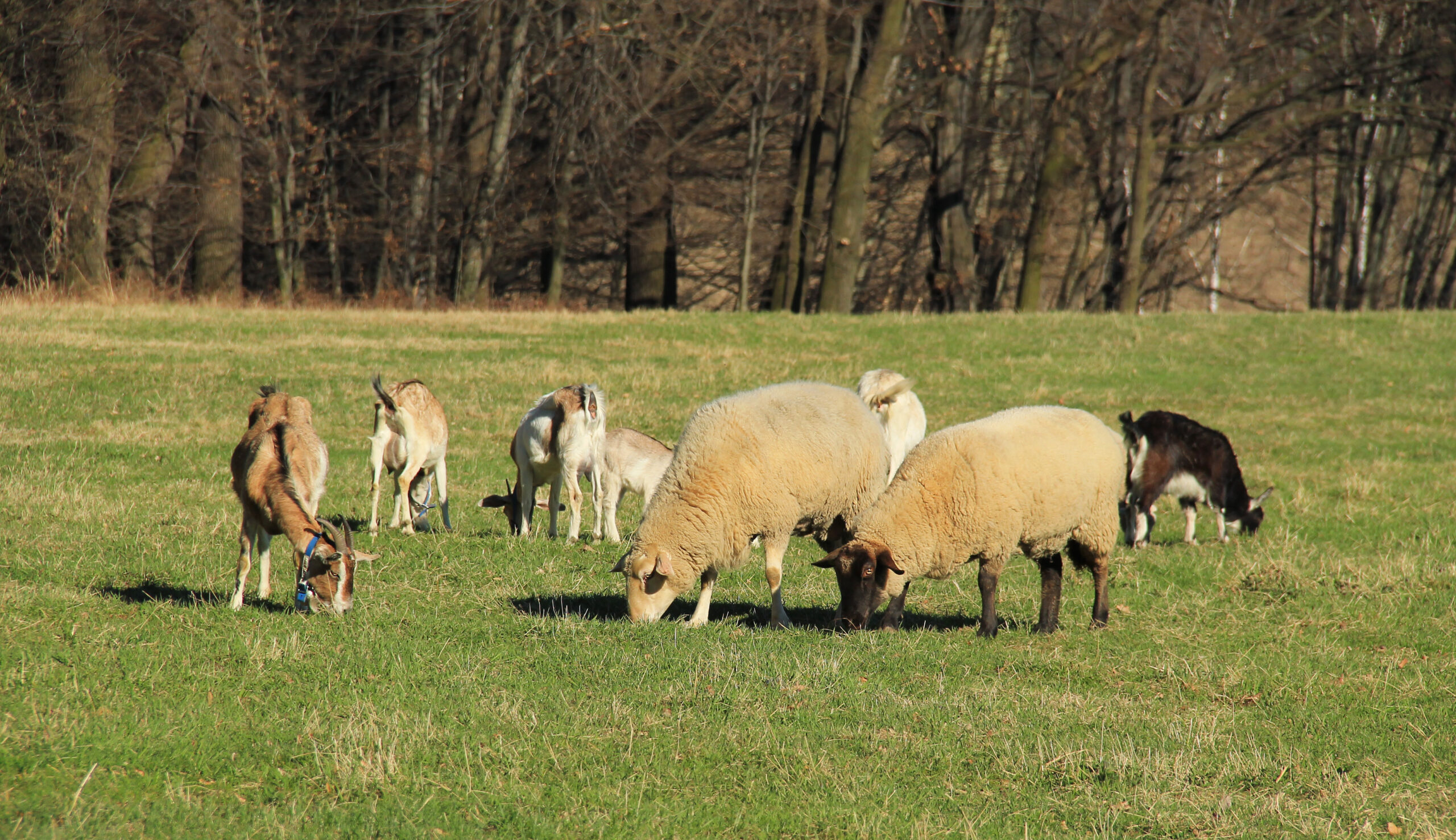
Scrapie is a chronic, progressive disease of the central nervous system that affects sheep and goats. Scrapie is the oldest of the group of neurodegenerative diseases known as transmissible spongiform encephalopathies (TSE). Some of the other TSE are Bovine Spongiform Encephalopathy known as mad cow disease, Chronic Wasting Disease which is found in deer, and Creutzfeldt Jacob Disease which is found in humans. TSE are protein-misfolding diseases that lead to brain damage and are always fatal.
The cause of Scrapie is not completely understood, but evidence indicates that an infectious protein referred to as a prion is responsible for the disease. These infectious prions cause damage to the normal prion proteins found in the brain. The mis-folding of the proteins lead to brain damage and the presentation of clinical signs of the disease. Prions are very resistant to destruction, so once in the environment, they are difficult to remove.
Scrapie is believed to primarily be transmitted by the oral route. Typically, lambs and kids might ingest the prion when they come in contact with the infectious agent through placentas and birthing fluids from infected ewes and does. Older animals may be exposed to the prions this way as well. Colostrum and milk are also sources of prions. Other secretions such as urine, feces, saliva, and nasal secretions may contain infectious prions as well. Once ingested, the prions cross into the lymphoid system. The prions will incubate for a long time usually two to five years before entering the nervous system.
Genetics plays a part in Scrapie infections. Certain breeds are more susceptible to the disease due to genetic composition. Genetic testing is available for producers to help them select breeding stock with resistant genes.
Clinical signs most commonly associated with Scrapie are intense pruritis, ataxia, and wasting. Early in the disease, small ruminant producers may notice slight changes in behavior with sheep and goats infected with Scrapie. Initially, animals may have a staring or fixed gaze, may not respond to herding, and may be aggressive towards objects. As the disease progresses, other clinical signs noticed are progressive weight loss with normal appetite, incoordination, head tremors, and intense pruritis. In the terminal stages, sheep are recumbent and may have blindness, seizures, and an inability to swallow. Once initial clinical signs are notice, death usually occurs in one to six months.
The gold standard for postmortem (dead animals) diagnosing of Scrapie is the use of immunohistochemistry test on brain tissues as well as microscopic examination of brain tissue for characteristic TGE lesions. Live animal diagnosis is possible by testing lymphoid tissues from the third eyelid and rectal mucosa scrapings.
There is no treatment available for Scrapie, so prevention is key to controlling the disease. Following biosecurity protocols is a good starting point for preventing Scrapie. Part of the biosecurity plan is to maintain a closed flock and only buy replacement animals from certified Scrapie free flocks. Producers should limit visitors’ contact with their animals. Sanitation is important in lambing and kidding areas. Manure and bedding contaminated with birthing fluids and placentas should be disposed of properly. Genetically resistant animals should be used for breeding to produce genetically resistant offspring.
It should be noted that there is a novel or atypical form of Scrapie. This disease may also be referred to as Nor98 variant. This atypical version of Scrapie was initially found in Norway. It has been diagnosed in the United States as well. The disease is usually only found in a single old animal in the flock or herd. The brain lesions in atypical Scrapie are different from classical Scrapie. Currently, experts believe that natural transmission of atypical Scrapie is not likely.
The United States Department of Agriculture (USDA) has been battling Scrapie for decades. According to recent information from the USDA, the United States (US) is close to accomplishing eradication of the disease. In order for the United States to achieve Scrapie free status, no sheep or goats can test positive for classical scrapie for seven years and a certain level of testing needs to be done each year that represents the sheep and goat populations within the country. Small ruminant producers can assist the USDA eradication efforts by contacting the USDA when they have an adult sheep or goat exhibiting clinical signs of Scrapie or an adult animal dies or is euthanized. Producers should contact the Oklahoma State Veterinarian, Dr. Rod Hall at 405-522-6141 or the USDA Veterinary Services at 405-254-1797. This will aid the USDA in reaching sampling testing goals. There is no charge for the collection or testing of the samples for scrapie.
Scrapie is a disease that needs to be eliminated from the US. Once eliminated, the US will have additional export markets for sheep and goat products. Oklahoma State University Cooperative Extension Service has an informative fact sheet on Scrapie. Please visit the Local County Extension Office and asked for fact sheet VTMD-9135 or producers may view the fact sheet online at https://extension.okstate.edu/fact-sheets/scrapie.html. Also, the USDA National Scrapie Eradication Program website has valuable information as well at https://www.aphis.usda.gov/aphis/ourfocus/animalhealth/animal-disease-information/sheep-and-goat-health/national-scrapie-eradication-program.
References Cassmann, E. D., & Greenlee, J. J. (2020). Pathogenesis, detection, and control of scrapie in sheep. American journal of veterinary research, 81(7), 600–614. https://doi.org/10.2460/ajvr.81.7.600





-
 Country Lifestyle2 years ago
July 2017 Profile: J.W. Hart
-
 Attractions9 years ago
48 Hours in Atoka Remembered
-
 Farm & Ranch2 years ago
From Plow to Plentiful: The Most Important Inventions in Agricultural History
-
 Equine9 years ago
Umbilical Hernia
-
 Outdoors8 years ago
Grazing Oklahoma: Honey Locust
-
 Country Lifestyle5 years ago
The Two Sides of Colten Jesse
-
 Farm & Ranch8 years ago
Hackberry (Celtis spp.)
-
 Equine6 years ago
On the Road with Emily Miller-Beisel