
Farm & Ranch
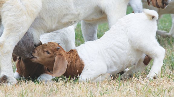
Johne’s Disease in Small Ruminants

By Barry Whitworth
Most small ruminant producers realize that the number one health problem in sheep and goats is gastrointestinal parasites (GIP) or what is commonly referred to as “worms.” Producers have several tools such as FAMACHA, Five Point Check, or Fecal Egg Counts that assist them in finding and treating animals infected with GIP.
These tools alert producers to compromised animals so that they can intervene with early treatment before the disease progresses too far. Still, on some occasions animals succumb to the “worms” and die. Many assume the treated animal died because the “worms” were resistant to the dewormer used to treat the parasites.
Sometimes this may be the case, but another possibility is that GIP was not the problem. Another disease that is sometimes mistaken for GIP is Johne’s disease. This disease has clinical signs that are very similar to GIP.
Johne’s disease, which is pronounced “yoh-nees,” is a gastrointestinal disease of ruminants that is fatal. The bacterium that causes the illness is Mycobacterium avium subspecies paratuberculosis which is commonly referred to by the acronym MAP. Producers should be aware that Johne’s is a herd disease. This means that if one animal is found ill with MAP, then several more of the normal appearing animals are probably also infected with the organism.
Most animals are infected early in life, but the clinical symptoms of the disease do not appear for many months. The organism is contagious. The bacterium can be transferred from animal to animal within a species such as sheep to sheep or from one ruminant to another ruminant such as cattle to sheep. Non-ruminant species (such as dogs, raccoons, coyotes, etc.) can be infected with MAP, but they usually to do not have any clinical signs. All breeds of sheep and goats are susceptible. Since very few records are kept, no one is sure how widespread the disease is in small ruminants.
MAP is very hardy in the environment. The organism is resistant to heat, cold, moisture, and drying. The bacterium has been found to survive for several months in the soil and water. The organism can be found in grasses fertilized with contaminated manure. Disinfectants that are labeled as “tuberculocidal” may be used for cleaning surfaces, feed troughs or water troughs. However, organic material may inactivate the disinfectant. Composting manure will reduce the number of MAP organisms.
MAP infects the last portion of the small intestines and its lymph nodes. This area is called the ileum. For reasons that have not been explained, the organism begins to replicate and infect more tissue. The immune system responds with inflammation. The ileum thickens, which prevents absorption of nutrients and ultimately results in the animal starving.
Learn more in the November issue!

Barry Whitworth, DVM
Area Food/Animal Quality and Health
Specialist for Eastern Oklahoma
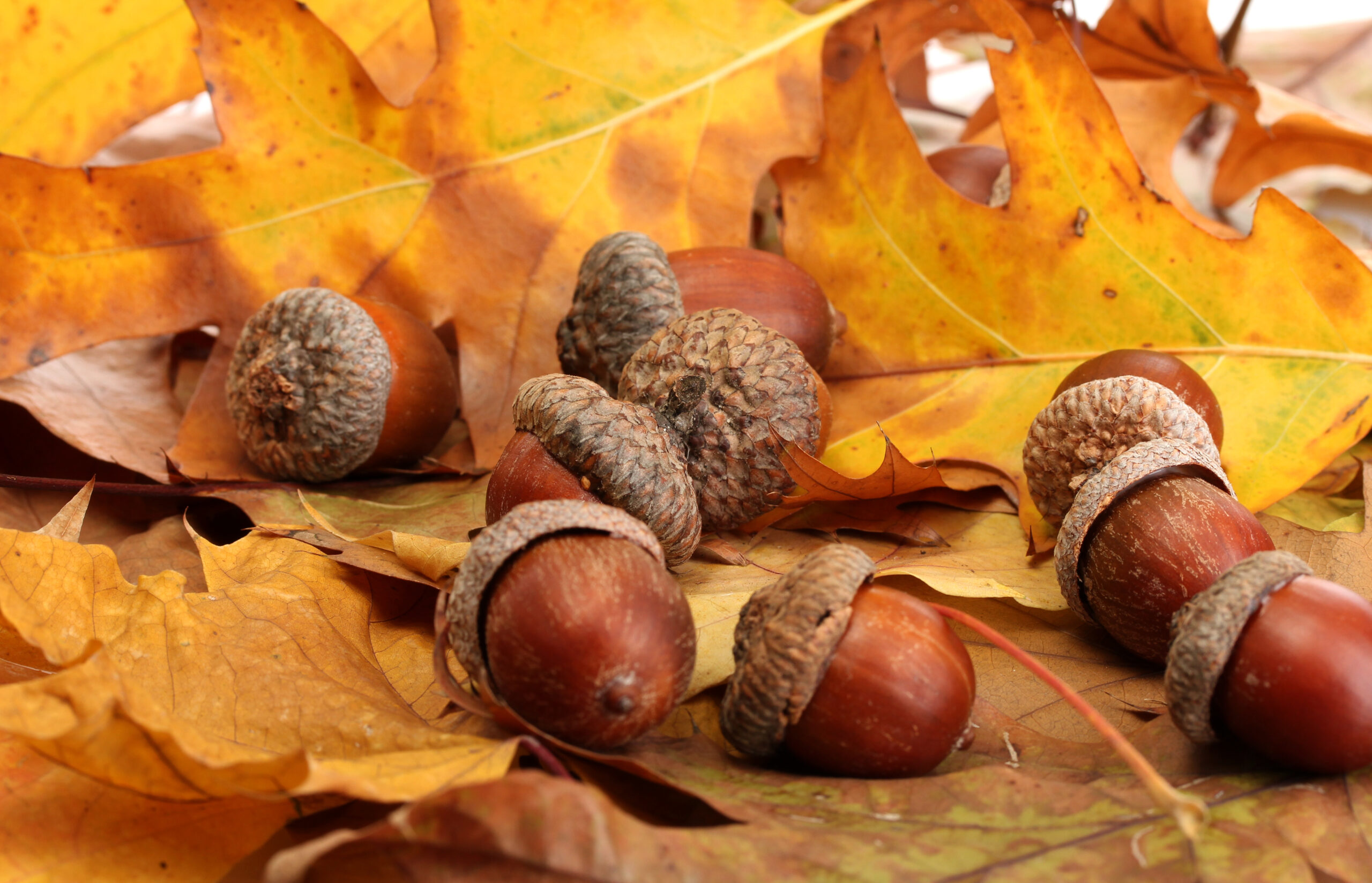
With the prolonged drought, most pastures in Oklahoma are in poor condition. With the lack of available forage, animals may go in search of alternative foods. If oak trees are in the pastures, acorns may be a favorite meal for some livestock this fall. This may result in oak poisoning.
Oak (Quercus species) leaves, twigs, buds, and acorns may be toxic to some animals when consumed. Obviously, acorns can be a problem in the fall and green acorns can be more toxic than mature acorns. When acorns form only a small portion of the diet, there are usually no signs of problems. However, consumption of large quantities may result in toxicity. Tannins in the acorns cause the toxicity. The most common tissue damaged by the tannins are the digestive tract and kidneys. Cattle and sheep appear to be more susceptible to toxicity than goats. Other animals such as horses, rabbits, and chickens have succumbed to the toxicity of oak poisoning as well. Interestingly, some individual animals are more tolerable of the toxins and show no ill effects when consuming acorns.
Clinical signs of oak toxicity usually appear a few days after consumption of acorns. Initially, the animals are weak, listless, emaciated, and anorexic. This is followed by ventral edema (swelling of lower parts of the body such as legs, chest, ventral abdomen), urinating large amounts of urine, abdominal pain, and constipation. The animal may pass hard mucus covered fecal material which may change to black tarry or bloody feces as the disease progresses. If the animal is not treated, kidney failure is likely.
A tentative diagnosis of acorn poisoning may be based on clinical signs and access to acorns. Blood tests that indicate kidney disease is another clue to the condition. A necroscopy with examination of tissues for characteristic lesions of the disease is the standard to confirm a diagnosis of oak toxicity.
Treatment of oak toxicity starts with removing the animals from the area where the acorns are located. Those animals displaying signs of the disease should be given fluids to correct dehydration and electrolyte imbalances. Mineral oil and/or activated charcoal may be given to reduce toxin absorption. If animals survive the initial toxicity, they may recover, but it may take several weeks for kidney function to return to normal.
As always, prevention is better than treatment. Producers should be very careful allowing livestock to graze in areas where acorns are present. Livestock should be fed plenty of hay and feed this fall to avoid over consumption of acorns. For those producers who cannot avoid grazing areas with large numbers of oak trees, feeding a grain mixture with 10% to 20% of calcium hydroxide has been successful in preventing problems with acorn poisoning.
Two thousand twenty-two has not been the best year for livestock producers. The drought has produced poor pasture conditions as well as very little hay. On top of those problems, feed costs continue to increase. The last problem a producer needs is a large number of sick cows. For those that graze an area with a large number of oak trees, prevention may be worth the cost this year. At the very least keep a close watch of your animals this fall. Producers wanting more information about oak toxicity, should consult with their local veterinarian or visit with their Oklahoma State University Cooperative Extension County Agriculture Educator.
Barry Whitworth, DVM | Area Food/Animal Quality and Health Specialist for Eastern Oklahoma
*Article originally printed in the October 2022 issue of Oklahoma Farm & Ranch.
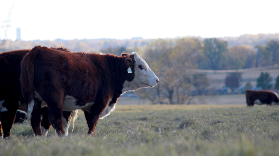
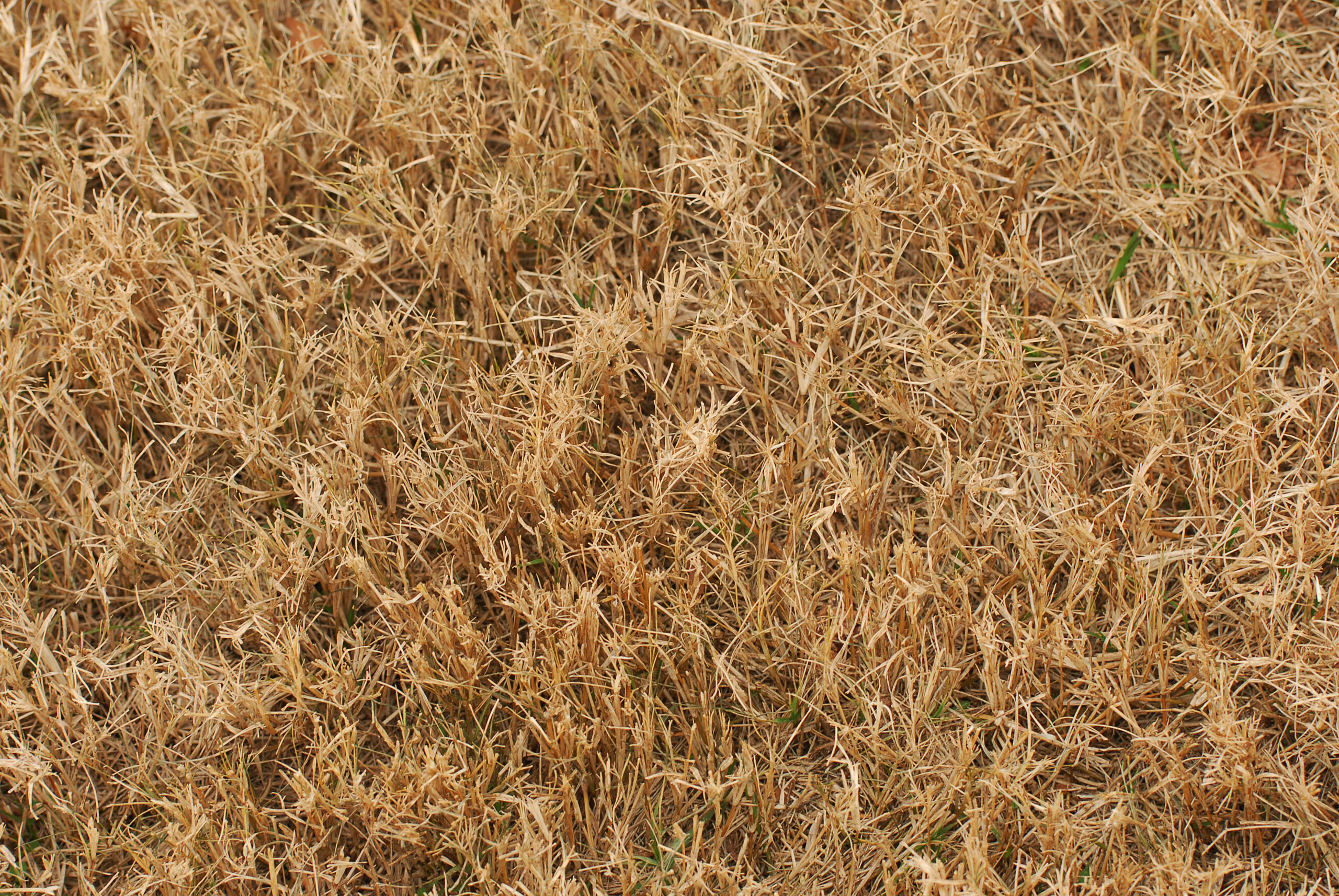
Since most of Oklahoma experienced drought conditions and with fall fast approaching, producers with fescue pastures should closely observe their livestock for any signs of fescue toxicity. According to Mike Trammel, Pottawatomie County Ag Educator and Muti-County Agronomist, fescue toxins (ergot alkaloids) tend to increase in Kentucky-31 tall fescue pastures in the fall. Some reports indicate more problems with fescue toxins following a summer drought and limited fall rains. All of this may put Oklahoma cattle at a greater risk of fescue toxicity.
One issue that cattle experience with fescue toxins is fescue foot. Fescue foot is thought to be caused by ergot alkaloids such as ergovaline. These alkaloids are produced by endophyte fungus (Epichloë coenophiala) which is in tall fescue. Ergovaline has been proven to be a vasoconstrictor which might be responsible for fescue foot and heat intolerance also known as summer slump in cattle. Other issues that may be seen with the ergot fescue toxins are reduced milk production and reproductive issues.
Clinical signs of fescue foot appear within a few days of cattle being turned on to tall fescue pastures or it may take weeks if toxins in the pasture are low. Producers will initially observe cattle with arched back, rough hair coats, and sore feet. These symptoms are more noticeable early in the morning and with cold weather. This is followed by reddening and swelling in the area between the dewclaws and hooves. The lameness usually becomes more severe with time. If no action is taken, gangrene will result in loss of tissues distal to the coronary band and declaws. If the weather remains mild, other signs such as increase respiration rate, increase heart rate, and higher body temperature are more common.
Other causes of lameness in cattle must be differentiated from fescue foot. One simple method that will help differentiate fescue foot from footrot is to check the temperature of the foot. If the foot is cold, this is an indication that the problem is more likely fescue foot.
Since there is not a specific treatment for fescue foot, the condition must be managed. Cattle need to be observed daily for any signs of lameness or stiffness during the first few weeks on fescue pastures. This should be done early in the morning before cattle walk off the stiffness. Producers should pay close attention during cold weather, especially when rain, snow, or ice are present. Any animal showing clinical signs of fescue foot should be removed from the pasture and placed in a clean environment. The animal should be fed a ration with no fescue toxins.
The best but most costly solution to reduce fescue toxicity is to renovate old pastures with new endophyte friendly varieties. If this option is not possible, producers might try interseeding fescue pastures with clovers or other grasses. This should dilute fescue toxins. Nitrogen fertilization may increase ergot alkaloids, so producers should avoid fertilizing fescue pastures with high amounts of nitrogen. Researchers have demonstrated that feeding a supplement while grazing fescue pastures reduces clinical symptoms. Some studies indicate a difference in susceptibility to fescue toxicity in some cattle. Selecting cattle based on genetic tolerance of fescue toxins is an option. (For more information go to www.agbotanica.com/t-snip.aspx)
With large areas in Oklahoma covered with Kentucky-31 fescue pastures, fescue foot as well as other fescue toxicities are not going away any time soon. Livestock producers will need to watch their livestock closely for any signs of fescue toxicity and manage their pastures to keep toxins as low as possible. If producers would like more information on fescue foot, they should consult their veterinarian and/or visit their local Oklahoma State University Cooperative County Extension Agriculture Educator.
By Barry Whitworth, DVM, MPH | Senior Extension Specialist Department of Animal & Food Sciences | Ferguson College of Agriculture | Oklahoma State University
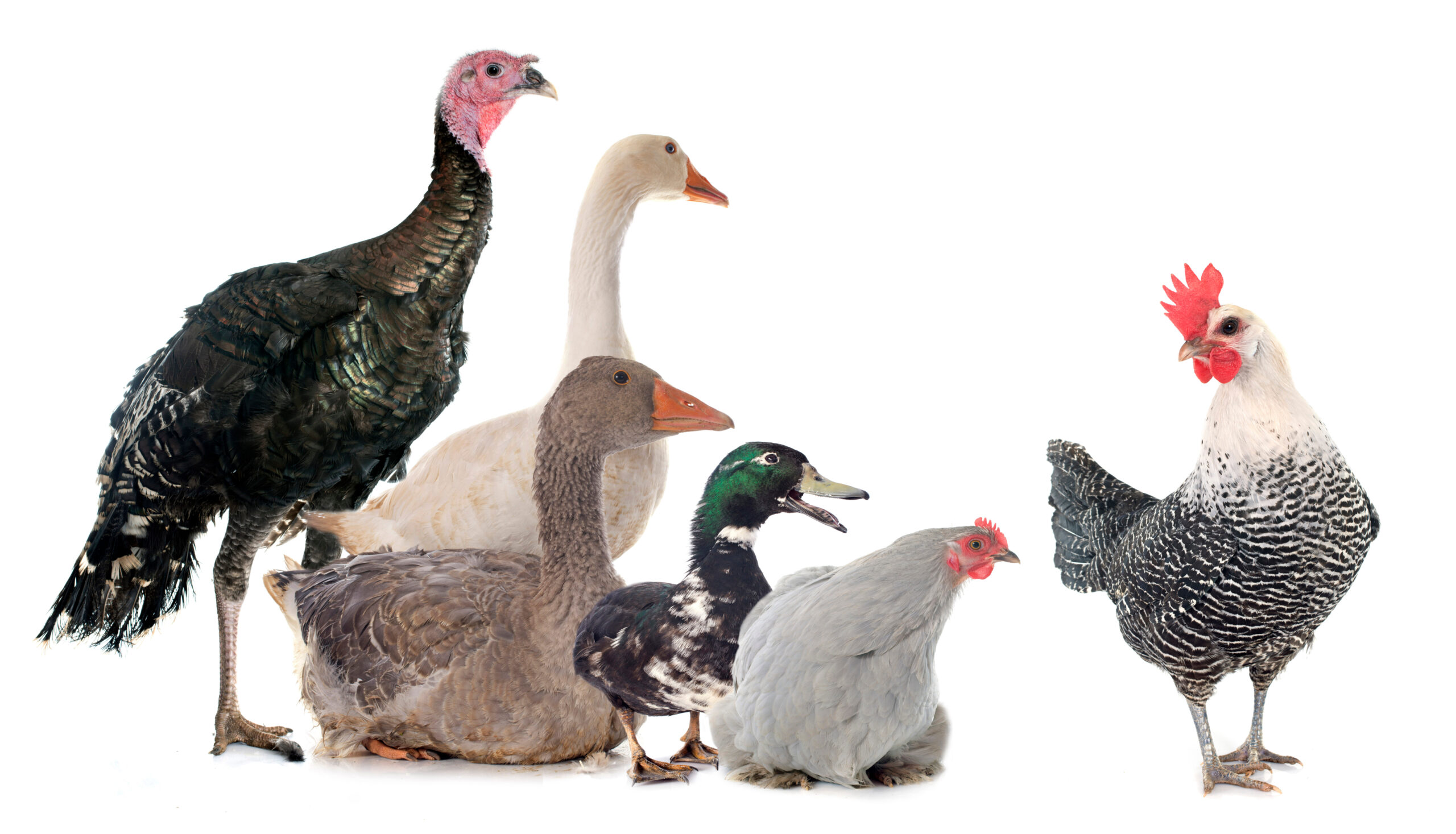
According to the 11th edition of Poultry Diseases, external parasites of poultry are arthropods that live on or in the skin and feathers. Essentially, parasites are freeloaders that live at the expense of the host. Backyard birds are infested with a variety of pests. Ticks, fleas, mites, and lice are some of the most common external parasites found in chickens, turkeys, and ducks. Several of these parasites are bloodsuckers. If not controlled, they can cause weight loss, decreased egg production, unthriftiness, and death in severe cases.
According to a study conducted by Dr. Amy Murillo and associates in California, the most common external parasites in backyard flocks were lice, fleas, and mites. Lice were the most frequently observed parasites, with the chicken body louse (Menacanthus stramineus) found on half of the premises inspected. The fluff louse (Goniocotes gallinae) was found in 35% of operations. The wing louse (Lipeurus caponis) and sticktight flea (Echidnophaga gallinacea) were present in 20% of flocks. Northern fowl mites (Ornithonyssus sylviarum), which are the most common mites found in commercial poultry operations, were detected in only 15% of flocks. However, the survey was conducted in the summer, which may have influenced the low number of northern fowl mites, since they are most active in the winter.
Birds infested with external parasites often become agitated due to skin irritation. They will spend more time preening and scratching. Their feathers may become damaged, and they may appear unhealthy. Birds showing these signs should be examined.
When examining birds for external parasites, producers should focus on the breast, back, head, vent region, and wings. Lice may be found on different parts of the body. They are yellowish in color and lie flat against the skin. Their eggs are typically found attached to the shafts of feathers. The vent area is the primary location to check for mite infestations and may appear “dirty.” Sticktight fleas are usually found embedded in the comb.
Birds should be monitored regularly. When producers are unable to examine all birds, they should focus on the young, the old, and any bird that appears unhealthy. The coop should also be inspected. Producers should examine the bedding, walls, and roosts, with close attention given to crevices and cracks where pests may hide.
Before parasite control can begin, the parasite must be correctly identified. Producers can use books or other publications for this purpose, or they may consult a veterinarian. Contacting the local Oklahoma State University Extension office is also a useful option. An agricultural extension educator may be able to identify the pest or submit samples to the Plant Disease and Insect Diagnostic Laboratory at Oklahoma State University for identification.
Prevention and control of external parasites require an integrated approach. The first line of defense is a strong biosecurity program to prevent parasites from entering the operation. Sanitation is also critical, keeping the coop and surrounding area clean helps prevent infestations.
Maintaining healthy birds is essential in preventing parasite infestations. Producers should focus on proper nutrition and disease prevention as they are key factors in maintaining a healthy flock. A strong immune system can help birds better withstand some external parasites.
Selecting the proper pesticide and using it correctly is essential. Many pests described in this article can be controlled with appropriate pesticides; however, their eggs are not killed, which requires repeated applications to target newly hatched larvae. Producers should read and follow pesticide label directions.
Alternative methods for external parasite control are also available such as providing diatomaceous earth mixed with sand for dust bathing or using sulfur bags to control mites and lice. For more information on these methods, see references below.
Finally, early identification and treatment greatly increase the chances of successful control. If infestations are allowed to become established, control becomes much more difficult.
For more information on external parasites in backyard poultry, producers may visit https://www.veterinaryentomology.org/ or contact their local veterinarian or Oklahoma State University County Agriculture Extension Educator.
References
Arends, J., J. (2003). External parasites and poultry pests. Diseases of Poultry. 11th Edition.
Murillo, A. C., & Mullens, B. A. (2016). Diversity and Prevalence of Ectoparasites on Backyard Chicken Flocks in California. Journal of medical entomology, 53(3), 707–71.
Murillo, A. C., & Mullens, B. A. (2016). Timing Diatomaceous Earth-Filled Dustbox Use for Management of Northern Fowl Mites (Acari: Macronyssidae) in Cage-Free Poultry Systems. Journal of economic entomology, 109(6), 2572–2579.
Murrillo, A.C., Mullens, B.A. (2016). Sulfur Dust Bag: A Novel Technique for Ectoparasite Control in Poultry Systems: Journal of Economic Entomology, 109(5), 2016, 2229-2233.





-
 Country Lifestyle2 years ago
July 2017 Profile: J.W. Hart
-
 Attractions9 years ago
48 Hours in Atoka Remembered
-
 Farm & Ranch2 years ago
From Plow to Plentiful: The Most Important Inventions in Agricultural History
-
 Equine9 years ago
Umbilical Hernia
-
 Country Lifestyle5 years ago
The Two Sides of Colten Jesse
-
 Outdoors8 years ago
Grazing Oklahoma: Honey Locust
-
 Farm & Ranch8 years ago
Hackberry (Celtis spp.)
-
 Equine6 years ago
On the Road with Emily Miller-Beisel