Equine
When a Nosebleed in Your Horse Means Business
It usually starts the same way. You walk into the barn, or pull your horse up after a hard run, and notice a streak of red sliding from one nostril. For most horse owners, that sight alone is enough to make your stomach drop. Nosebleeds in horses — known medically as epistaxis — can be anything from a harmless trickle to a serious medical emergency. The trick is knowing the difference.
Understanding What’s Really Going On
“Epistaxis” simply means bleeding from the nostrils. It can look dramatic, especially on a horse’s light-colored muzzle, but not every case is cause for panic. The real question is where the blood is coming from and why it started. Horses can bleed from several different parts of their upper and lower respiratory systems, ranging from the delicate nasal passages at the tip of the nose to deep within the lungs
External signs don’t always tell the full story. A steady stream of blood might come from a relatively minor nasal scrape, while a single drop could signal a deeper issue if it occurs repeatedly. Because of that, determining the origin of the bleed often requires a veterinarian’s examination and, in some cases, diagnostic tools like endoscopy or radiographs.
Still, horse owners can gather useful clues before the vet arrives. What was your horse doing just before the nosebleed began? Did it happen after intense exercise or while the horse was standing quietly in the stall? Is blood coming from one nostril or both? Has this happened before — and if so, always from the same side? Observations like these help narrow down the list of possible causes.
If blood is merely dripping or running slowly, chances are you’re not dealing with an immediate emergency. But if it’s flowing freely — more like a faucet than a leak — or doesn’t stop within a few minutes, it’s time to call your veterinarian.
Common Causes of Equine Nosebleeds
The list of potential sources for a horse’s nosebleed is long, but they generally fall into a few categories.
One of the most common and least serious is nasal mucosal trauma — a simple scrape or irritation of the tissues lining the nasal passage. Horses are curious creatures and not always careful about what they bump into. A playful nose rub on a rough fence board or an overly enthusiastic sneeze can rupture a tiny blood vessel and cause a short-lived trickle of blood. Passing a nasogastric tube or removing a foreign body can also irritate the area temporarily.
A more persistent cause is progressive ethmoid hematoma, a vascular mass that forms within the nasal cavity or sinuses. These growths often bleed intermittently and almost always from the same nostril. The bleeding is usually modest but tends to recur over time. While the initial episodes may not look alarming, the mass will continue to grow if untreated, so early veterinary intervention gives the best chance of successful removal.
Another possibility involves the sinuses themselves. Trauma, infection, or even small fractures to the skull can lead to bleeding within the sinus cavities. Horses are notorious for finding new and inventive ways to injure themselves — banging into doors, slipping in the trailer, or catching a halter just wrong — and sometimes the only outward sign is a slow bleed from the nose. Small fractures often heal with rest, but significant ones may require surgical repair.
Bleeding from both nostrils usually suggests a deeper origin. One of the best-known examples is Exercise-Induced Pulmonary Hemorrhage (EIPH), sometimes called “bleeding” in racehorses. This occurs when capillaries within the lungs rupture under the extreme pressure of intense exercise, sending blood up through the trachea and out both nostrils. Horses affected by EIPH might cough, swallow repeatedly, or show decreased performance after a run. Though it’s most common in racehorses, it can appear in any equine athlete pushed to their limits.
The most dangerous cause of all is guttural pouch mycosis, a fungal infection of the guttural pouches — air-filled sacs located behind the horse’s skull that connect to major arteries. The fungus, often Aspergillus fumigatus, thrives in warm, dark, and moist environments such as hay and soil. As it grows, it erodes the arterial walls, sometimes silently, until a vessel ruptures and the horse begins to bleed heavily from the nose. In some cases, this can lead to fatal blood loss within minutes. If a horse shows even minor, unexplained nosebleeds that repeat from the same side, it’s worth scheduling an endoscopic exam to rule this out.
When It’s Time to Act
It’s not always easy to tell which kind of nosebleed you’re dealing with, but the following general rule applies: the more rapid and continuous the bleeding, the more urgent the situation. If the blood is coming in spurts, pooling quickly, or refuses to stop, treat it as an emergency. A horse can lose a significant amount of blood in a short period, especially if a major artery is involved.
On the other hand, a small amount of blood that stops on its own and doesn’t return likely points to a minor issue. Even so, it’s smart to keep notes — which nostril bled, how long it lasted, what the horse was doing, and any other symptoms you noticed. This information can help your veterinarian determine whether further testing is necessary.
If your horse experiences recurring nosebleeds, particularly from the same side, don’t ignore them. Repetition can be the biggest red flag of all.
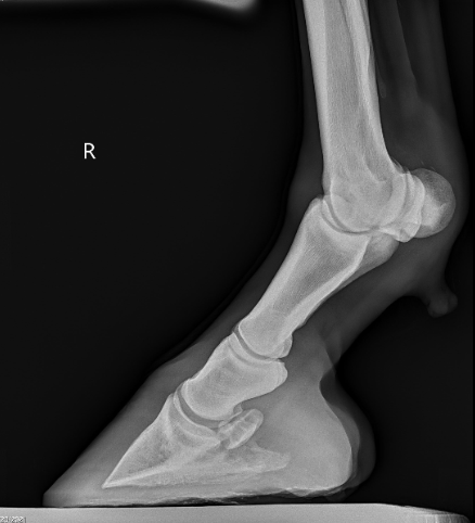
When you call your vet, expect a methodical evaluation. They’ll first perform a physical exam and may use an endoscope, a flexible camera designed to navigate the nasal passages and visualize the sinuses, guttural pouches, and upper airway. Endoscopy allows the veterinarian to identify the exact source of bleeding — whether a scraped mucosa, a growing hematoma, or a fungal lesion.
If there’s evidence of trauma, radiographs may be taken to assess bone integrity. Horses with suspected guttural pouch infections or ethmoid hematomas often undergo additional imaging or even surgical procedures to address the underlying issue. For suspected EIPH, an endoscopic exam performed within an hour or two after exercise can confirm blood in the trachea and lungs.
While you’re waiting for the veterinarian, keep your horse calm and still. Excitement or movement can raise blood pressure and worsen bleeding. Remove tack, halter, or anything that might interfere with breathing. Resist the urge to pack or plug the nostrils — this can cause more harm than good. Instead, observe closely and document what you see. If safe to do so, a quick photo or short video can be helpful for your vet later.
Do not tilt the horse’s head upward, as that can allow blood to flow backward into the airway. Let the horse lower its head naturally. Fresh air and a quiet space are best until professional help arrives.
A horse’s nosebleed can stop your heart for a moment, but it doesn’t always mean disaster. Many are brief and harmless, the result of a bump or sneeze in just the wrong way. But others — particularly those that are heavy, prolonged, or recurring — can point to serious underlying disease.
When in doubt, treat every nosebleed as something that deserves attention. Take a deep breath, make careful observations, and get your veterinarian involved early. In the long run, those few extra minutes of vigilance can make all the difference.
References
“Equine Epistaxis: What You Need to Know.” The Horse, American Association of Equine Practitioners.
“Exercise-Induced Pulmonary Hemorrhage in Horses.” Merck Veterinary Manual.
“Nosebleeds in Horses — When Do You Need to Be Concerned?” Horse & Hound.
“Epistaxis (Nosebleed) in Horses.” PetMD.
“Nasal Hemorrhage in the Horse: Where and Why.” DVM360 Proceedings.
By Dr. Garrett Metcalf
The suspensory ligament is a vital component of the limb of a horse to produce normal locomotion and support. The suspensory ligament is a common area of concern in performance horses of various disciplines and can be single handedly the cause of lameness or performance issues. This article is going to look at a specific degenerative disease of the suspensory ligament and what horses are at risk for this disease.
DSLD or degenerative suspensory ligament desmitis was first discovered in the early 1980’s in Peruvian Paso horses. The name has been changed because the suspensory ligament is not the only organ affected from the disease but the suspensory is ultimately the biggest issue. The newer name, ESPA or equine systemic proteoglycan accumulation, is more correct because other ligaments and tissues are affected by this disease. In this article we will only focus on the suspensory ligament. The most commonly affected breeds are Peruvian Paso, Paso Fino, Morgan, Saddlebred, Warmblood, Paints, American Quarter Horse, and Thoroughbred breeds. The age of onset of the disease is variable among breeds but it is more common to be seen in middle age to older horses. However it has been documented in horses as young as one year of age. The disease generally will have a slow insidious onset that can go undiagnosed for months or years depending on the horses work and discipline.
A horse that begins to show early signs of DSLD may have a vague lameness issue that is difficult to isolate and they most likely will resolve with a period of rest. As the horse returns to moderate level of work the lameness will return. This scenario may go on for several months or more before the discovery of the DSLD is made. The first indication of DSLD is often pain isolated in the suspensory branches or fetlock region when a flexion test is performed. Horses with DSLD will also have a “dropped” fetlock appearance because the suspensory is the main supporting structure of the fetlock joint. DSLD can affect the hind limbs, forelimbs or all limbs at the same time. A unique sign of DSLD is that not just one limb is affected but rather bilaterally affecting the limbs, meaning it will either affect either both forelimbs or hind limbs at the same time. It is my experience that the hind limbs are more commonly affected compared to the forelimbs. Horses will often have enlargement of the fetlock region and increased joint fluid or wind puffs. Horses will often have a straight hock or post legged hind limb appearance. Horses will often shift weight frequently in an effort to get relief from the discomfort and this can be confused with other lameness issues or foot related pain.
Diagnosis of DSLD is often made by clinical signs, breed and ultrasound findings. Ultrasound imaging of the suspensory ligaments will often show diffuse enlargement of the suspensory body and branches. The suspensory ligament will have a poor heterogeneous fiber pattern with periligamentious soft issue thickening from scar tissue deposition and edema or fluid within the tissue. Radiographs of the lower limb may reveal abnormal bone changes in the sesamoid bones behind the fetlock joints and even osteoarthritis of the pastern and or fetlock joints. A definitive diagnosis can be made from a biopsy of a ligament in the neck called the nuchal ligament, but is not often performed because of the invasiveness of the biopsy.
Treatment is very limited and it is mostly geared towards protection of further damage by prolonged rest. Pain management is also important to attempt to keep the horse as comfortable as possible. Different shoeing techniques can be used with marginal success. In early cases of DSLD, a suspensory shoe that helps engage more work from the deep digital flexor tendon can help elevate the fetlock and offer more protection to the suspensory ligament. The devastating thing about this disease is that there is no cure and there are hardly any good options to slow the progression of the disease. DSLD carries a poor prognosis when the diagnosis is made in any breed of horse or any discipline. Although some cases can be managed better than others, it often progresses to the point of debilitating pain and discomfort to the point of humane euthanasia especially in the Peruvian Paso breed.
Read more in the February 2023 issue of Oklahoma Farm & Ranch.
By Dr. Devan England DVM
Does your horse have gastric ulcers? Gastric or stomach ulcers are frequently blamed for a variety of things including poor performance, acting ‘cinchy’, weight loss, not eating, poor coat condition, diarrhea and colic. However, gastric ulcers are not always the culprit and the only way to know for sure if your horse has gastric ulcers is to look at the stomach on camera, using an endoscope. Poor appetite and poor body condition are the mostly widely observed clinical signs with gastric ulcers, however, these are non-specific. If you think your horse might have gastric ulcers, the best place to start is to talk to your veterinarian and consider scheduling a gastroscopy. Gastroscopy requires the horse be held off feed for at least 16-18 hours and held off water for at least 6-8 hours. Fasting off feed and water is necessary to allow the veterinarian to see the whole stomach. If restricting feed or water is difficult in your management situation, many veterinarians will allow you to hospitalize your horse the night before gastroscopy for proper fasting.
Gastric ulcers are split into two types, classified by the location of the ulcer in the stomach. Squamous ulcers are ulcers that occur in the squamous or skin like portion of the stomach. This is the top part of the horse’s stomach, is closest to the esophagus, and has squamous tissue to protect this portion of the stomach from stomach acids. The other ulcer type are glandular ulcers. Glandular ulcers occur in the bottom portion of the stomach, which is closest to the small intestine. This portion of the stomach has glandular mucosa with cells responsible for producing stomach acids for digestion as well as cells that produce mucus and buffers to protect the lining from stomach acid. Gastroscopy is important not only for diagnosing whether ulcers are present but also determining the severity and the type of ulcer, because these two ulcer types require different treatments.
Squamous gastric ulcers are common in racehorses both in and out of training, with higher prevalence in racehorses under training. Prevalence in Thoroughbred racehorses in training has been reported to be up to 100% (Sykes 2015). Squamous ulcers are also prevalent in Western pleasure horses, Thoroughbred stallions on breeding farms, and Italian donkeys (Sykes 2015). Glandular gastric ulcer prevalence has not been as well described as squamous ulcers. Glandular ulcers are reported to be most common in Thoroughbred and Standardbred racehorses, Canadian showjumpers and polo ponies, and American Quarter Horses (Sykes 2015).
Risk factors for ulcers vary by ulcer type. Anti-inflammatories (Bute, Banamine) can increase the risk of glandular ulcers in some horses by affecting normal defense mechanisms but are not a high risk in most horses. Horses that display stereotypic behaviors, such as cribbing, have an increased risk of squamous ulcers. Grain fed before hay in non-exercising horses, feeding larger amounts of grain, and increased time between meals increases the risk of squamous ulcers. Increased time with high intensity exercise and housing in single pens is associated with increased risk of glandular ulcers. A straw only diet, lack of water access and lack of direct contact with other horses increases the general risk of gastric ulcers.
If your horse is diagnosed with ulcers, the mainstay of treatment is a buffered formulation of omeprazole (Gastrogard, Ulcergard). Over the counter Omeprazole and compounded Omeprazole are not effective because without buffering, the acidic stomach quickly breaks down the drug before absorption. Most horses with squamous ulcers will have healing of these ulcers after a 4-week course of Gastrogard or Ulcergard at treatment dose (whole tube for the average horse). Some horses may be healed by 3 weeks of treatment, but all horses should undergo a recheck gastroscopy before stopping treatment. Horses diagnosed with glandular ulcers need combination therapy with Gastrogard/Ulcergard and Sucralfate for 4 weeks. About 2/3 of horses with glandular ulcers will heal in this time, but some horses may require longer treatment times so a recheck is always recommended before discontinuing treatment.
Horses at higher risk of gastric ulcers may benefit from preventative (low) doses of Ulcergard (1/4 tube in average sized horse) given for a few days before and during high stress situations like long distance travel and competitions. Sea buckthorn berry supplement may be protective against formation of glandular ulcers. Dietary management to decrease the risk of ulcers includes providing more frequent small hay meals if pasture access is not available, limiting high sugar grains as much as possible and adding vegetable oil to the feed.
Sykes BW, Hewetson M, Hepburn RJ, Luthersson N, Tamzali Y. European college of equine internal medicine consensus statement – equine gastric ulcer syndrome in adult horses. J Vet Internal Med 2015; 29:1288-1299.
By Janis Blackwell
As the season arrives to gear up for participation in your equine event of choice, one thing remains a constant for all horse owners. That constant is our responsibility to insure the safety of our horses by being diligent to maintain the integrity of the trailers in which we haul them. There are a number of things that can be dangerous both inside and outside of your trailer. Whether you traveled all winter long or whether your trailer sat unused or was used very little through the cold weather months, at least once a year your trailer is due a thorough going over. So here we go with a checklist that will help you insure a happy and safe trip for you and your equine partner.
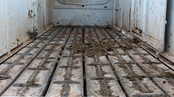
- A sound floor is absolutely imperative. Whether your floor is aluminum, steel or wood, it should be cleaned regularly after use to preserve it. Urine and manure will erode and weaken all types of floors. Even rubber mats will not prevent erosion of your floor. (Maintenance tip: remove mats and wash aluminum floors often to prevent erosion.)
- Especially check wooden floors for rotten boards. Immediately replace questionable flooring before hauling. (Maintenance tip: For wood, remove mats and wash out manure and debris. Coat wooden floor in a cheap motor oil. Allow to sit in hot summer weather until the oil soaks in. Be careful—floor will be slippery until oil cures into the wood. This treatment yearly will preserve a wooden floor for much longer than normal as it repels urine and protects the wood).
- Keep the interior and exterior washed to enable you to check for rusted out places, leaks, etc.
- Have a professional check your brakes at least once yearly to be sure they are operating properly.
- Be sure tires are inflated to the proper air pressure, and check the inside of each tire for hidden unusual wear that could cause a blowout. Replace worn tires before leaving home.
- Wheel bearings must be checked and packed at least once a year. This should be done even if the trailer has been rarely used since the last time the wheel bearings were packed. In fact, trailer maintenance professionals say that sitting stationary and unused is even worse for the bearings. Improper care and maintenance of wheel bearings can cause a wheel to seize up and actually twist off while in use. Use a horse trailer professional for this maintenance task.
- Axles should be checked for bowing. A bent or bowed axle can cause excessive tire wear and damage wheel bearings.
- There should be no more than two inches in height difference from the front of the trailer to the back. More difference than that causes the bulk of weight of the trailer and its contents to ride mostly on the rear axle causing it to bow and wear on both tires and wheel bearings.
- Another critical part of the trailer to keep an eye on are the butt chain or bar and the back door. The butt chain or bar should be firmly attached to the wall and its keeper and should always be latched. The door should have a strong secure latch with a pin to insure it stays latched while in motion.
- Finally, but certainly not of least importance is a thorough check of the trailer hitch including ball and coupling. Keep the ball well greased. Periodically, check to see that the ball is still securely tightened and the latch on the coupling is working properly.
These few critical safety check points can save you money, stress and the wellbeing of your horse. Until next time, happy trails and safe traveling.
This article was originally published in the April 2016 issue of Oklahoma Farm & Ranch.


-
 Country Lifestyle2 years ago
July 2017 Profile: J.W. Hart
-
 Attractions9 years ago
48 Hours in Atoka Remembered
-
 Equine9 years ago
Umbilical Hernia
-
 Outdoors8 years ago
Grazing Oklahoma: Honey Locust
-
 Country Lifestyle4 years ago
The Two Sides of Colten Jesse
-
 Farm & Ranch1 year ago
From Plow to Plentiful: The Most Important Inventions in Agricultural History
-
 Farm & Ranch8 years ago
Hackberry (Celtis spp.)
-
 Equine6 years ago
On the Road with Emily Miller-Beisel