Equine
History of the Horseshoe Part 2
By Laci Jones
With the saying, “Don’t reinvent the wheel,” Lee Liles, owner of the National Museum of Horse Shoeing Tools and Hall of Honor said the same could be said about the horseshoe.
“Nothing is really new in a horseshoe,” he explained. “It just seems to be a revolving circle.”
While the horseshoe has not changed, Liles said the title of the horseshoe specialist has. At some point over time, the horseshoer took on the name “farrier,” he added.
Certification
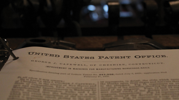
Horseshoers in the late 19th century needed to have a certificate issued by the Master Horseshoers National Protective Association. Liles said the association was like a union. If someone was caught without being a licensed member, they could be fined $400.
The color of the certificates was different each year, making the certificates easy to identify when entering the blacksmith’s shop.
Other countries like Canada and Germany also required certification. Horseshoers in London in 1909 needed to carry a certificate with them at all times. The certificate was signed by the queen, Liles explained.
Mid-Century Trends
In the 1940s, 1950s and 1960s, all horses had heel caulks on their horseshoes.
“I can remember back in the 1960s we had a shoe we called ‘The Cowboy Shoe,’” he added. The Cowboy Shoe was beveled out on the inside to help shed dirt. Liles said a good cow pony on rough terrain requires a tougher shoe.
“If it weren’t for a horseshoer moving up [in rough country] there would never have been good cattle country,” he said. “He kept the horses sound from being on the rocks.”
He remembered when owners could get away with just trimming their ranch horses in the 1960s. “Getting into the 1970s, you had to put shoes on ranch horses,” Liles added. “Their feet weren’t as strong because the breeding changed their feet a lot.”
The Horse
“The life of a horseshoer is very short,” Liles explained, “especially this day and time more so than the old days.”
To give a time-life-history of the horse, 24 million horses were in the United States in 1915. By 1950, they were down to two million. Farmers began working with tractors and the workhorse phased out, he added. In the late 1950s and early 1960s, horses became luxury items, Liles explained. Owners used horses more on ranches and started horse shows and events.
“In 1965, there was a shortage of horseshoers because most were cavalry horseshoers or taught by somebody who was in the cavalry,” he added. Those horseshoers retired by the 1960s, and there was a need for horseshoeing schools.
Z-Bar Shoe
“Our shoe industry has changed dramatically since 1979,” Liles explained. “We’ve got so may good horseshoes on the market today that you hardly need a forge to heat them up to shape them.”
Liles said the industry now has front and rears in the patterns, which has changed horseshoeing. The Z-Bar shoe may have been patented in 1900 in New Orleans, but the shoe has become more popular in recent years. “It’s popular in our horseshoeing contests,” Liles explained.
The Z-Bar shoe is used on horses that have a bad quarter crack, he added. The shoe relieves the pressure and lets the swelling go down, and the bar distributes the weight in the foot. This is the only patent I ever seen south of the Mason-Dixon Line,” he added.
Current Trends
“Just in the last few years, we’re seeing horseshoes with multiple nail holes,” Liles added. “That is more than we’ve ever had.”
For 200 years in the United States, horseshoes have always had eight nail holes in the shoe. Now, 10 to 16 nail holes are in a shoe. “That’s a dramatic change,” Liles explained. “It gives [farriers] more of an option to where they can rotate the nails around if the foot gets bad, so it’s not a bad deal.”
Shoeing horses on the front end and not the back end is trending in the show horse industry, he explained. When asked why this is trending, Liles said one person started winning, and everybody started thinking it would help them win.
“It’s not always necessary to shoe the back end of a horse,” he said. “Most of the weight on a horse is carried from the front end with the weight of the horse’s neck and the cowboy.”
Marvin Beeman, DVM and educator for the American Colt Horse Association, told Liles how a horse farrier shoes a ranch horse depends on the biographical areas in which they live in.
“If you change your horse from one environment to another environment, it will actually change the growth pattern of the horse’s foot and start a groove around his foot,” Liles explained. “When you change that horse’s environment, the horseshoer can actually read it in his foot.
“That’s hard for a lot of people to imagine, but when you bring a horse from the east coast to the west coast, that’s a dramatic change for that horse.”
Different Materials
Liles said using different materials is a current trend among horse farriers. In the early days of horseshoeing, farriers used wrought iron. Today, most horseshoes are made using plain steel. However, the show horse industry uses a lot of aluminum and titanium.
“A pair of heavy walking horse shoes can cost $5,000,” Liles explained, “but it’s made out of tungsten.”
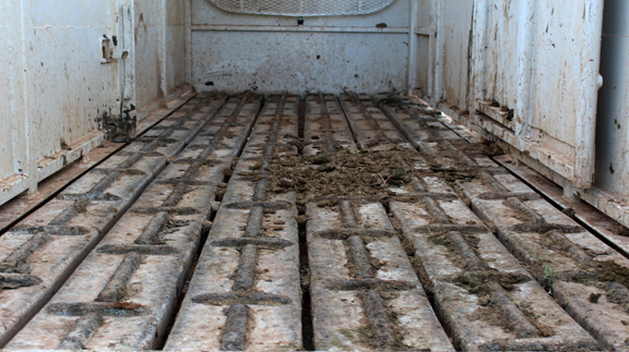
Plastic and rubber shoes have also become popular in recent years. Amish horses used on roads have horseshoes made of Borium. Borium is a texture that keeps the shoe from wearing out, but it can also have a negative impact on the road.
“If you drive around in Amish or Mennonite country and see a dip in the road, it’s from a horse going down the road,” Liles explained. “A lot of states like Pennsylvania and Ohio have funded projects trying to come up with a traction device horseshoe that will not hurt their asphalt and highways as bad.”
Glue-on shoes have come along after he was an active horse farrier. The dairy industry uses the glue-on shoes more than anybody, he added. Cattle are on water and concrete, and they can have a tremendous problem with foot rot. Liles said proper nutrition can help combat this disease.
“I’ve never seen many horse’s feet that I couldn’t nail a shoe on,” Liles said, “but, I see where they help a lot of horses today.”
To learn more about the history of the horseshoe, visit www.horseshoeingmuseum.com.
This article was originally posted in the August 2016 issue of Oklahoma Farm & Ranch.
By Summer McMillen
Everyone that knows anything about horses knows that there are bad ones, good ones, and great ones.
The bad ones are good for nothing. You can’t catch them, you can’t saddle them, and you can’t get on them without feeling like you need a helmet, some kind of padded vest and, an instruction manual. Once you do mount up the whole ride is a battle and heaven forbid, you actually have a job to do because they are little to no help in holding the herd. We all find ourselves owning a bad one or two throughout our lives. Best case scenario is they find a more tolerable home to go to through via a horse sale or the classifieds. Worst case scenario all you can do is say “Vaya con Dios,” put a sign on them that reads “Do Not Attempt,” and turn them out to pasture. Hoping they are decent enough to stay within the borders and make a beautiful yard ornament.
Good horses are usually much more tolerable. They’re pretty easy to catch, saddle, and hop up on. Sometimes they might have a bad habit or two like setting back when they’re tied to a fence or, getting cold backed on early mornings that you tolerate because they are so skilled in a specific field. A good horse is usually only good for one thing. They have a niche talent m, if you will. They can be a good heel horse. A good head horse. The horse you want to gather pastures on because you know he won’t knicker or rare up when you get dropped off in the jig line. A good kid horse. Your rodeo horse. The horse you put your wife on when she’s being a little wimpy that day. Good horses usually get sold because they are proficient in their given field and they find good homes making both parties happy. We will all own many good horses in our lives and be happy to do so.
Great horses are a rare and treasured possession. They are simultaneously easy and hard to own. Easy because you can do anything on them. Hard because everyone is always trying to buy them from you. A great horse stands still while your kid pulls their head down all the way to the ground so they can halter them. A great horse is never cold backed and always ready to cinch tight and take off. A great horse can be ridden in the pasture and the rodeo arena on the same day. A great horse doesn’t need practice. A great horse is always willing to do anything you ask of them at any given moment. Great horses find their homes as horse colts and usually live out the rest of their days at the same home because great horses are irreplaceable.
People and horses are not all that different. There are bad, good, and great ones. The more time I spend around horses the more I am convinced of the kind of person I want to be. “Bad” will absolutely not do. “Good“ is much too common and just doesn’t quite cut it more often than not. “Great” is what I aspire to be.
Great can be defined in so many ways when we let human standards get involved but, I want to be great as defined in the qualities of a great horses.
I want to be kind and patient while my children are learning. I want to be ready to help anyone who asks me. I want to go the extra mile. I want to make my home a beautiful place to come to after a full day’s work outside. I want to not be thrown off by life’s twists and turns but, firm in my faith.
So, basically what I’m saying is I want to be a great horse. And honestly there are worse things we could all aspire to be.
Here’s to great horses. May we know them, love them, and if we’re lucky be great just like them.
By Dr. Garrett Metcalf
The suspensory ligament is a vital component of the limb of a horse to produce normal locomotion and support. The suspensory ligament is a common area of concern in performance horses of various disciplines and can be single handedly the cause of lameness or performance issues. This article is going to look at a specific degenerative disease of the suspensory ligament and what horses are at risk for this disease.
DSLD or degenerative suspensory ligament desmitis was first discovered in the early 1980’s in Peruvian Paso horses. The name has been changed because the suspensory ligament is not the only organ affected from the disease but the suspensory is ultimately the biggest issue. The newer name, ESPA or equine systemic proteoglycan accumulation, is more correct because other ligaments and tissues are affected by this disease. In this article we will only focus on the suspensory ligament. The most commonly affected breeds are Peruvian Paso, Paso Fino, Morgan, Saddlebred, Warmblood, Paints, American Quarter Horse, and Thoroughbred breeds. The age of onset of the disease is variable among breeds but it is more common to be seen in middle age to older horses. However it has been documented in horses as young as one year of age. The disease generally will have a slow insidious onset that can go undiagnosed for months or years depending on the horses work and discipline.
A horse that begins to show early signs of DSLD may have a vague lameness issue that is difficult to isolate and they most likely will resolve with a period of rest. As the horse returns to moderate level of work the lameness will return. This scenario may go on for several months or more before the discovery of the DSLD is made. The first indication of DSLD is often pain isolated in the suspensory branches or fetlock region when a flexion test is performed. Horses with DSLD will also have a “dropped” fetlock appearance because the suspensory is the main supporting structure of the fetlock joint. DSLD can affect the hind limbs, forelimbs or all limbs at the same time. A unique sign of DSLD is that not just one limb is affected but rather bilaterally affecting the limbs, meaning it will either affect either both forelimbs or hind limbs at the same time. It is my experience that the hind limbs are more commonly affected compared to the forelimbs. Horses will often have enlargement of the fetlock region and increased joint fluid or wind puffs. Horses will often have a straight hock or post legged hind limb appearance. Horses will often shift weight frequently in an effort to get relief from the discomfort and this can be confused with other lameness issues or foot related pain.
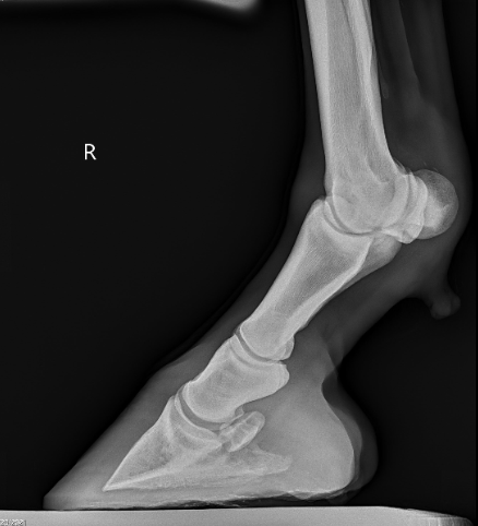
Diagnosis of DSLD is often made by clinical signs, breed and ultrasound findings. Ultrasound imaging of the suspensory ligaments will often show diffuse enlargement of the suspensory body and branches. The suspensory ligament will have a poor heterogeneous fiber pattern with periligamentious soft issue thickening from scar tissue deposition and edema or fluid within the tissue. Radiographs of the lower limb may reveal abnormal bone changes in the sesamoid bones behind the fetlock joints and even osteoarthritis of the pastern and or fetlock joints. A definitive diagnosis can be made from a biopsy of a ligament in the neck called the nuchal ligament, but is not often performed because of the invasiveness of the biopsy.
Treatment is very limited and it is mostly geared towards protection of further damage by prolonged rest. Pain management is also important to attempt to keep the horse as comfortable as possible. Different shoeing techniques can be used with marginal success. In early cases of DSLD, a suspensory shoe that helps engage more work from the deep digital flexor tendon can help elevate the fetlock and offer more protection to the suspensory ligament. The devastating thing about this disease is that there is no cure and there are hardly any good options to slow the progression of the disease. DSLD carries a poor prognosis when the diagnosis is made in any breed of horse or any discipline. Although some cases can be managed better than others, it often progresses to the point of debilitating pain and discomfort to the point of humane euthanasia especially in the Peruvian Paso breed.
Read more in the February 2023 issue of Oklahoma Farm & Ranch.
By Dr. Devan England DVM
Does your horse have gastric ulcers? Gastric or stomach ulcers are frequently blamed for a variety of things including poor performance, acting ‘cinchy’, weight loss, not eating, poor coat condition, diarrhea and colic. However, gastric ulcers are not always the culprit and the only way to know for sure if your horse has gastric ulcers is to look at the stomach on camera, using an endoscope. Poor appetite and poor body condition are the mostly widely observed clinical signs with gastric ulcers, however, these are non-specific. If you think your horse might have gastric ulcers, the best place to start is to talk to your veterinarian and consider scheduling a gastroscopy. Gastroscopy requires the horse be held off feed for at least 16-18 hours and held off water for at least 6-8 hours. Fasting off feed and water is necessary to allow the veterinarian to see the whole stomach. If restricting feed or water is difficult in your management situation, many veterinarians will allow you to hospitalize your horse the night before gastroscopy for proper fasting.
Gastric ulcers are split into two types, classified by the location of the ulcer in the stomach. Squamous ulcers are ulcers that occur in the squamous or skin like portion of the stomach. This is the top part of the horse’s stomach, is closest to the esophagus, and has squamous tissue to protect this portion of the stomach from stomach acids. The other ulcer type are glandular ulcers. Glandular ulcers occur in the bottom portion of the stomach, which is closest to the small intestine. This portion of the stomach has glandular mucosa with cells responsible for producing stomach acids for digestion as well as cells that produce mucus and buffers to protect the lining from stomach acid. Gastroscopy is important not only for diagnosing whether ulcers are present but also determining the severity and the type of ulcer, because these two ulcer types require different treatments.
Squamous gastric ulcers are common in racehorses both in and out of training, with higher prevalence in racehorses under training. Prevalence in Thoroughbred racehorses in training has been reported to be up to 100% (Sykes 2015). Squamous ulcers are also prevalent in Western pleasure horses, Thoroughbred stallions on breeding farms, and Italian donkeys (Sykes 2015). Glandular gastric ulcer prevalence has not been as well described as squamous ulcers. Glandular ulcers are reported to be most common in Thoroughbred and Standardbred racehorses, Canadian showjumpers and polo ponies, and American Quarter Horses (Sykes 2015).
Risk factors for ulcers vary by ulcer type. Anti-inflammatories (Bute, Banamine) can increase the risk of glandular ulcers in some horses by affecting normal defense mechanisms but are not a high risk in most horses. Horses that display stereotypic behaviors, such as cribbing, have an increased risk of squamous ulcers. Grain fed before hay in non-exercising horses, feeding larger amounts of grain, and increased time between meals increases the risk of squamous ulcers. Increased time with high intensity exercise and housing in single pens is associated with increased risk of glandular ulcers. A straw only diet, lack of water access and lack of direct contact with other horses increases the general risk of gastric ulcers.
If your horse is diagnosed with ulcers, the mainstay of treatment is a buffered formulation of omeprazole (Gastrogard, Ulcergard). Over the counter Omeprazole and compounded Omeprazole are not effective because without buffering, the acidic stomach quickly breaks down the drug before absorption. Most horses with squamous ulcers will have healing of these ulcers after a 4-week course of Gastrogard or Ulcergard at treatment dose (whole tube for the average horse). Some horses may be healed by 3 weeks of treatment, but all horses should undergo a recheck gastroscopy before stopping treatment. Horses diagnosed with glandular ulcers need combination therapy with Gastrogard/Ulcergard and Sucralfate for 4 weeks. About 2/3 of horses with glandular ulcers will heal in this time, but some horses may require longer treatment times so a recheck is always recommended before discontinuing treatment.
Horses at higher risk of gastric ulcers may benefit from preventative (low) doses of Ulcergard (1/4 tube in average sized horse) given for a few days before and during high stress situations like long distance travel and competitions. Sea buckthorn berry supplement may be protective against formation of glandular ulcers. Dietary management to decrease the risk of ulcers includes providing more frequent small hay meals if pasture access is not available, limiting high sugar grains as much as possible and adding vegetable oil to the feed.
Sykes BW, Hewetson M, Hepburn RJ, Luthersson N, Tamzali Y. European college of equine internal medicine consensus statement – equine gastric ulcer syndrome in adult horses. J Vet Internal Med 2015; 29:1288-1299.


-
 Country Lifestyle2 years ago
July 2017 Profile: J.W. Hart
-
 Attractions9 years ago
48 Hours in Atoka Remembered
-
 Equine9 years ago
Umbilical Hernia
-
 Farm & Ranch1 year ago
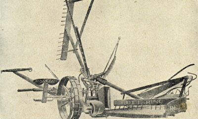
From Plow to Plentiful: The Most Important Inventions in Agricultural History
-
 Outdoors8 years ago
Grazing Oklahoma: Honey Locust
-
 Country Lifestyle5 years ago
The Two Sides of Colten Jesse
-
 Farm & Ranch8 years ago
Hackberry (Celtis spp.)
-
 Equine6 years ago
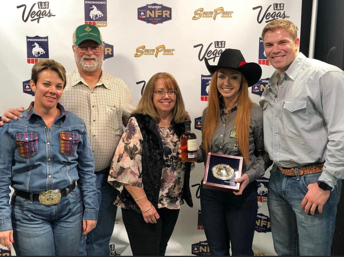
On the Road with Emily Miller-Beisel